U.S. flag

An official website of the United States government

Dot gov

The .gov means it's official.
Federal government websites often end in .gov or .mil. Before sharing sensitive information, make sure you're on a federal government site.

Https

The site is secure.
The https:// ensures that you are connecting to the official website and that any information you provide is encrypted and transmitted securely.

Skip to Main Navigation
Skip to Page Content
Skip to Atlas Navigation

Kidney - Mineralization

Image of mineralization in the kidney from a male F344/N rat in an acute study
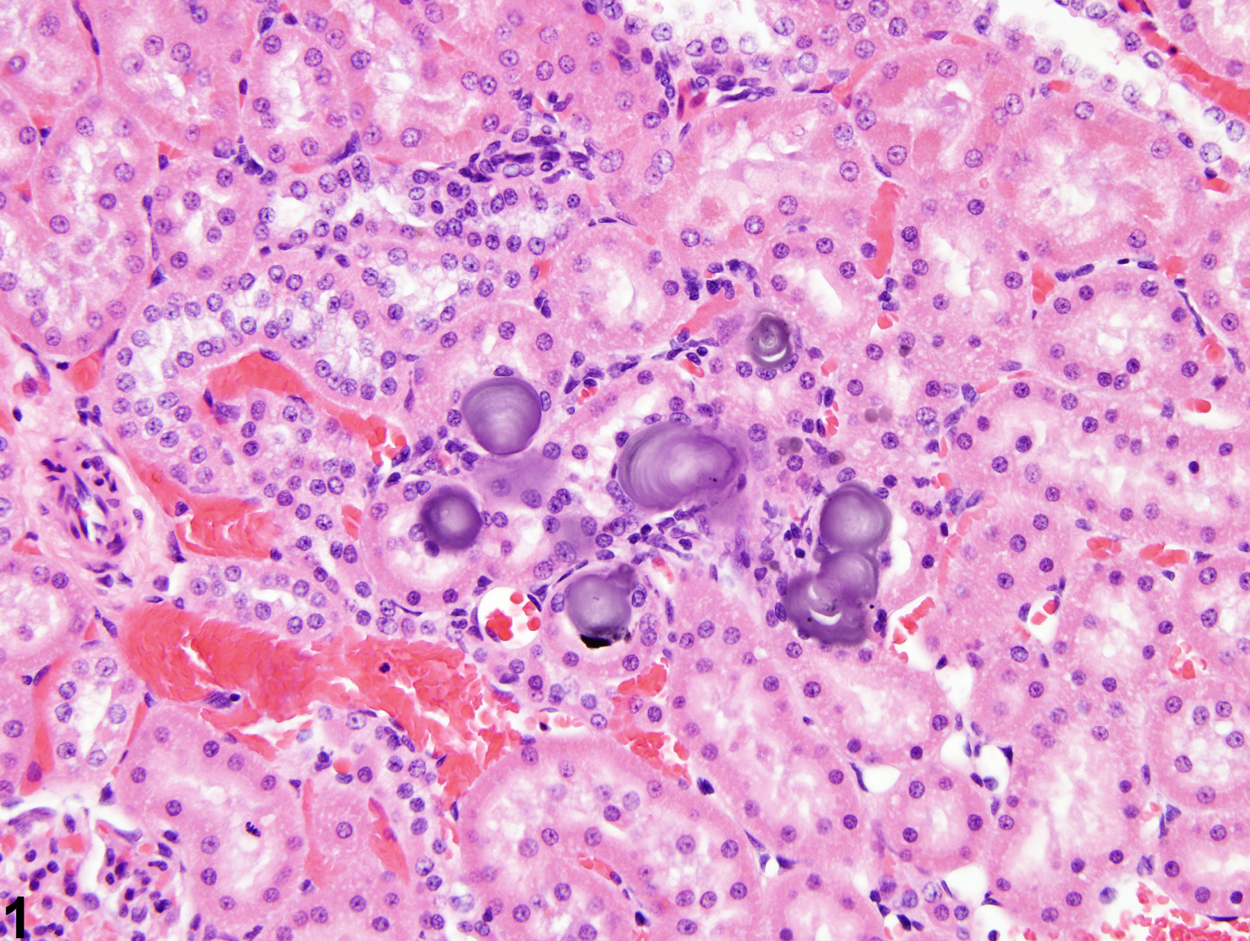
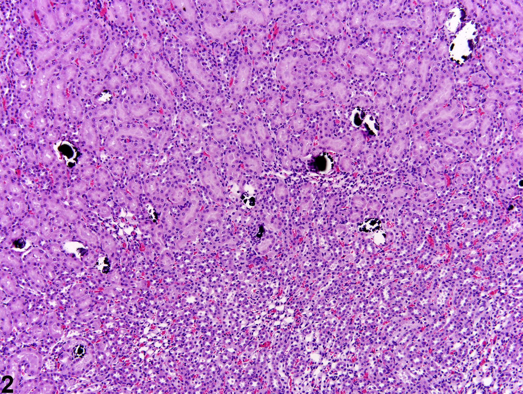
Kidney - Mineralization in a male F344 rat from an acute study. Spontaneous mineralization of cortical tubules is present.
Figure 1 of 3
Image of mineralization in the kidney from a female Harlan Sprague-Dawley rat in an acute study
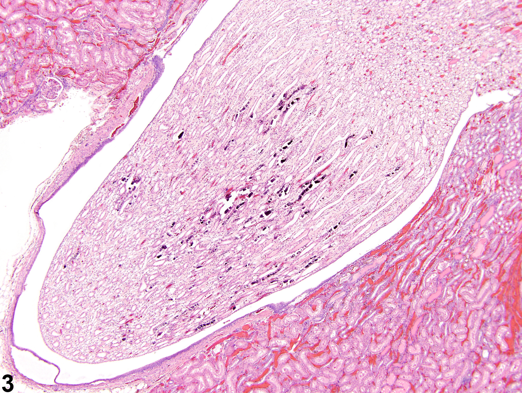
Kidney - Mineralization in a female Sprague-Dawley rat from an acute study. Mineralization is commonly observed in the area of the outer stripe and inner stripe of the outer medulla.
Figure 2 of 3
Image of mineralization in the kidney from a male F344/N rat in a chronic study
Kidney - Mineralization in a male F344/N rat from a chronic study. Linear mineralization of papillary tubules is associated with alpha-2u-globulin nephropathy.
Figure 3 of 3
next arrow

comment:

Mineralization is more commonly associated with spontaneous and minute background findings of basophilic deposits in the renal cortex, medulla,or papilla of rats and mice ( Figure 1image opens in a pop-up window ). In general, these depositshave no pathologic significance. However, mineralization may also be seen as a consequence to degeneration and necrosis. Mineralization may be induced by chemicals, hormones, or diet. Mineralized deposits usually involve tubule epithelia but may also involve the interstitium. Alizarin red S or von Kossa’s special stains are used to demonstrate mineral in histologic sections. Mineralized deposits seen particularly in female rats are noted in the pars recta and thin loops of Henle near the junction of the outer stripe of the outer medulla and the inner stripe of the outer medulla ( Figure 2image opens in a pop-up window ). Linear deposits of papillary mineralization are characteristic of chronic chemical administration that induces α2u-globulin nephropathy ( Figure 3image opens in a pop-up window ). Artifactual mineralization is sometimes encountered in the outer cortex. The cause of this mineralization often cannot be identified.

recommendation:

Mineralization should be diagnosed and graded. If small deposits of focal mineralization are recognized as a spontaneous background finding, they need not be diagnosed and the pathologist should use his or her judgment in deciding whether or not they are prominent enough to warrant diagnosis. When diagnosed, the pattern of the mineralization (e.g., linear papillary mineralization, focal medullary mineralization) should be described in the pathology narrative.

references:

Gopinath C, Prentice DE, Lewis DJ. 1987. Atlas of Experimental Toxicological Pathology. MTP Press, Lancaster, UK, 77-88.

Ritskes-Hoitinga J, Beynen AC. 1992. Nephrocalcinosis in the rat: A literature review. Prog Food Nutr Sci 16:85-124.
Abstract: http://www.ncbi.nlm.nih.gov/pubmed/1620876