U.S. flag

An official website of the United States government

Dot gov

The .gov means it's official.
Federal government websites often end in .gov or .mil. Before sharing sensitive information, make sure you're on a federal government site.

Https

The site is secure.
The https:// ensures that you are connecting to the official website and that any information you provide is encrypted and transmitted securely.

Skip to Main Navigation
Skip to Page Content
Skip to Atlas Navigation

Kidney - Nephroblastematosis

Image of nephroblastematosis in the kidney from a female Harlan Sprague-Dawley rat in a subchronic study
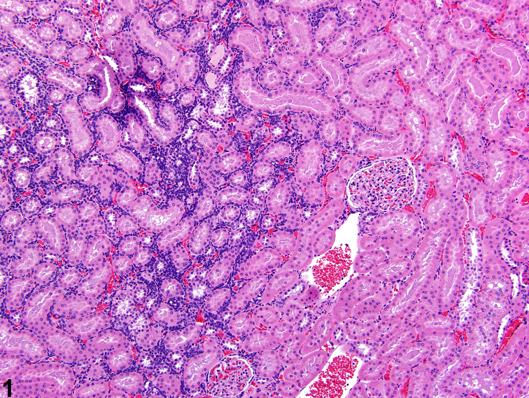
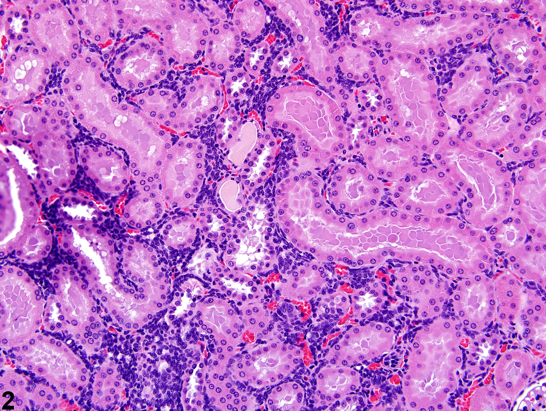
Kidney - Nephroblastematosis in a female Harlan Sprague-Dawley rat from a subchronic study. An area of dense basophilic cellularity can be seen in the renal cortex.
Figure 1 of 4
Image of nephroblastematosis in the kidney from a female Harlan Sprague-Dawley rat in a subchronic study
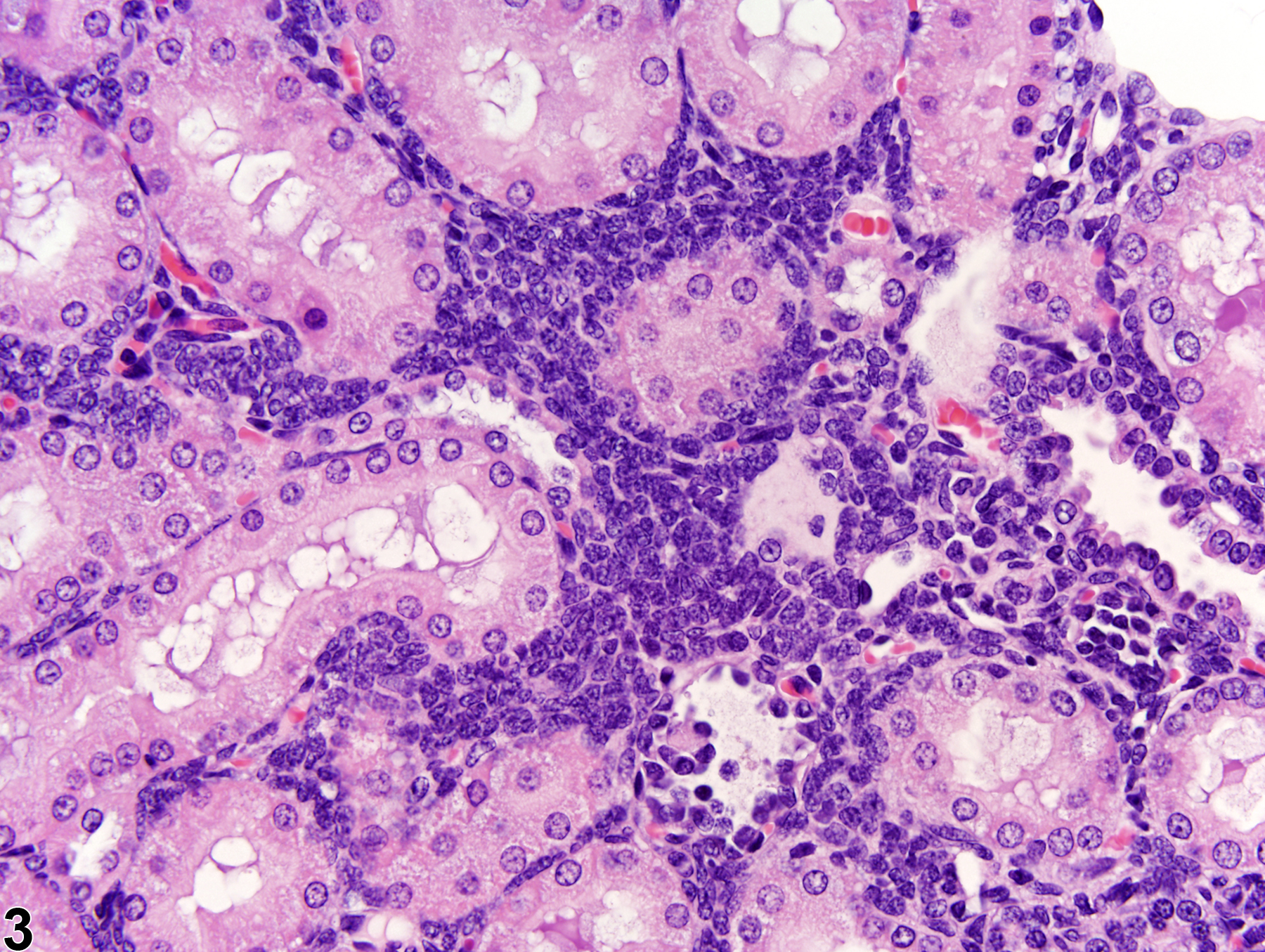
Kidney - Nephroblastematosis in a female Harlan Sprague-Dawley rat from a subchronic study. The cells are deeply basophilic and uniform in morphology. They are infiltrating between renal tubules, and no organoid differentiation of the lesion can be recognized.
Figure 2 of 4
Image of nephroblastematosis in the kidney from a female Harlan Sprague-Dawley rat in a subchronic study
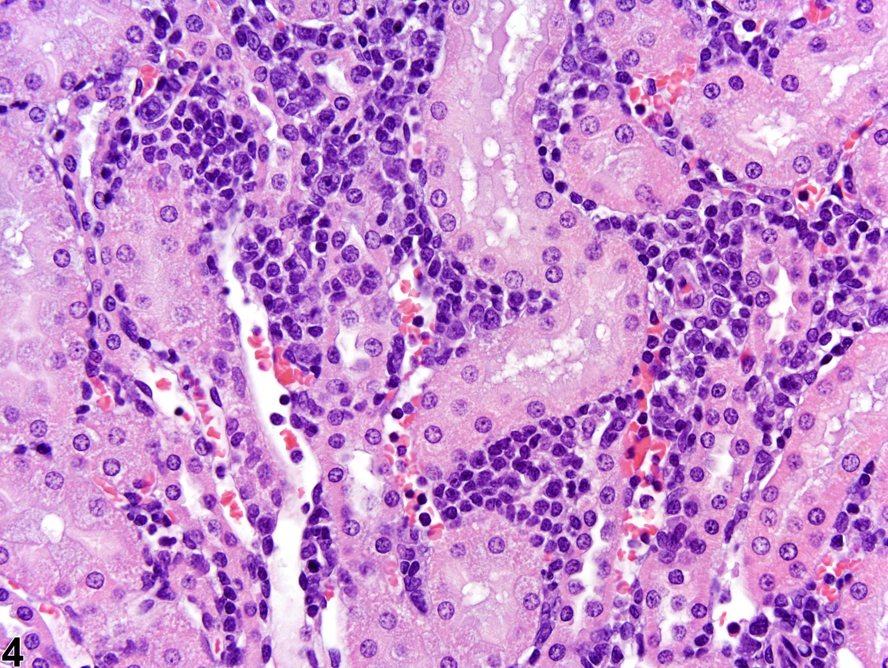
Kidney - Nephroblastematosis in a female Harlan Sprague-Dawley rat from a subchronic study. At higher magnification, the cells in nephroblastematosis have indistinct cell margins and cytoplasm, round to oval nuclei with stippled chromatin, and often a prominent nucleolus.
Figure 3 of 4
Image of nephroblastematosis in the kidney from a female Harlan Sprague-Dawley rat in a subchronic study
Kidney - Infiltration, Cellular, Mononuclear in a female Harlan Sprague-Dawley rat from a subchronic study. This higher magnification of mononuclear cell infiltration, characterized by a heterogeneous population of cells in the kidney and small blood vessels, is for comparison with Figures 1 - 3.
Figure 4 of 4
next arrow

comment:

Nephroblastematosis can be a spontaneous lesion and is the presumptive precursor lesion of nephroblastoma. On low power, nephroblastematosis appears as an area of densely basophilic cellularity ( Figure 1image opens in a pop-up window and Figure 2image opens in a pop-up window ). On higher magnification, uniform cells are seen infiltrating between renal tubules and are characterized by having indistinct cell margins and cytoplasm and round to oval nuclei with stippled chromatin ( Figure 3image opens in a pop-up window ). The distinction between nephroblastematosis and nephroblastoma is somewhat arbitrary and depends on differentiation into any organoid (tubule, glomeruloid) structures, which is seen in nephroblastematosis, and the overall size of the lesion. Differentiation from other lesions such as cellular infiltrates, inflammation, or lymphoma usually depends on the presence of a more heterogeneous population of cell types, which often have a focal to multifocal perivascular distribution, and/or the presence of associated cells in blood vessels within the lesion ( Figure 4image opens in a pop-up window ).

recommendation:

Nephroblastematosis should be diagnosed whenever present and given a severity grade.

references:

Frazier KS, Seely JC, Hard GC, Betton G, Burnett R, Nakatsuji S, Nishikawa A, Durchfeld-Meyer B, Bube A. 2012. Proliferative and non-proliferative lesions in the rat and mouse urinary system. Toxicol Pathol 40:14S-86S.
Abstract: http://www.ncbi.nlm.nih.gov/pubmed/22637735

Mesfin GM. 1999. Intralobar nephroblastemosis: Precursor lesion of nephroblastoma in the Sprague-Dawley rat. Vet Pathol 36:379-390.
Abstract: http://www.ncbi.nlm.nih.gov/pubmed/10490205