

Urinary System
Kidney, Papilla - Necrosis
Narrative
Brix AE. 2002. Renal papillary necrosis. Toxicol Pathol 30:672-674.
Abstract: http://www.ncbi.nlm.nih.gov/pubmed/12512867Frazier KS, Seely JC, Hard GC, Betton G, Burnett R, Nakatsuji S, Nishikawa A, Durchfeld-Meyer B, Bube A. 2012. Proliferative and non-proliferative lesions in the rat and mouse urinary system. Toxicol Pathol 40:14S-86S.
Abstract: http://www.ncbi.nlm.nih.gov/pubmed/22637735Greaves P. 2012. Histopathology of Preclinical Toxicity Studies, 4th ed. Elsevier, Amsterdam, 574-576.
Abstract: http://www.sciencedirect.com/science/book/9780444538567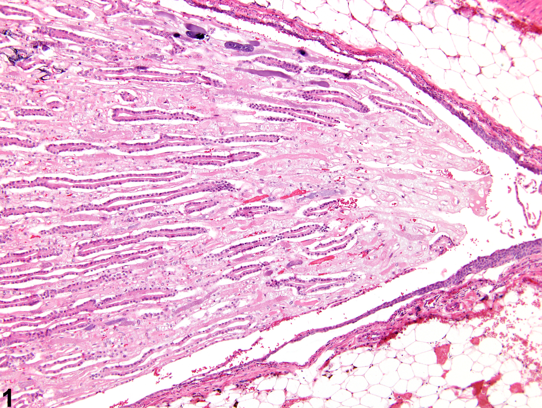
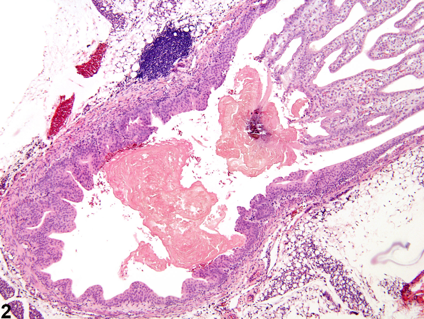
Kidney, Papilla - Necrosis in a male F344/N rat from a chronic study. Early necrosis of the papilla shows loss of cellular detail and staining affinity.
All Images

Kidney, Papilla - Necrosis in a male F344/N rat from a chronic study. Early necrosis of the papilla shows loss of cellular detail and staining affinity.

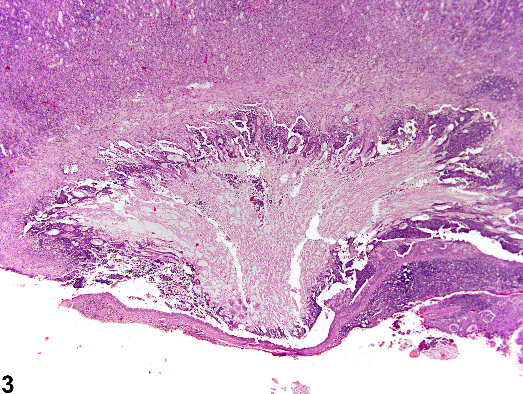
Kidney, Papilla - Necrosis in a male F344/N rat from a subchronic study. A portion of the necrotic renal papilla is sloughed off into the renal pelvis.

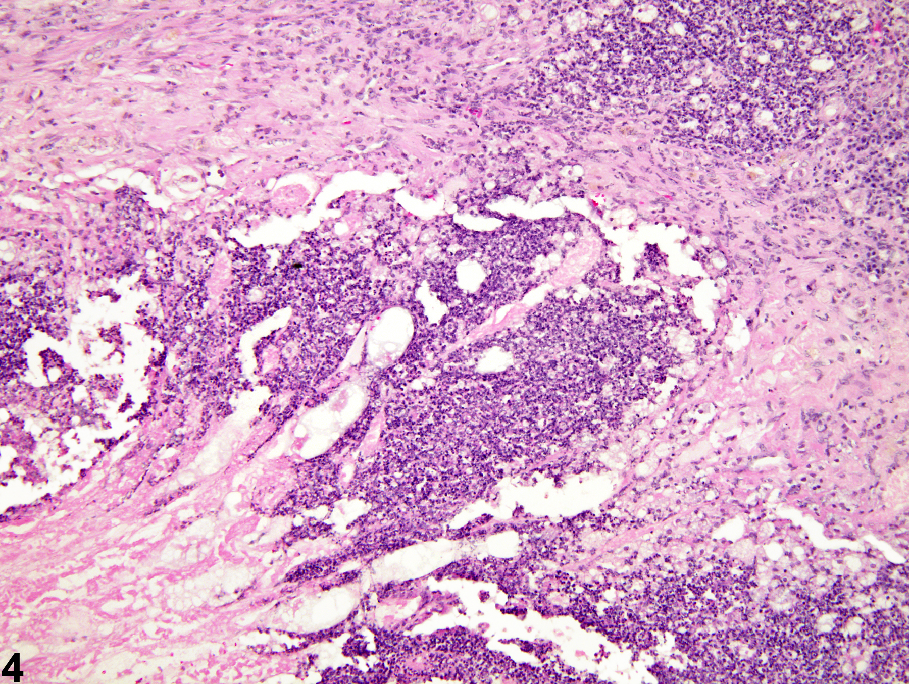
Kidney, Papilla - Necrosis in a female B6C3F1 mouse from a chronic study. There is a large area of papillary necrosis with a prominent peripheral zone of inflammation.

Kidney, Papilla - Necrosis in a male B6C3F1 mouse from a chronic study (higher magnification of Figure 3). Acute necrosis and inflammatory infiltrate are present.