

Urinary System
Kidney, Renal Tubule - Dilation
Narrative
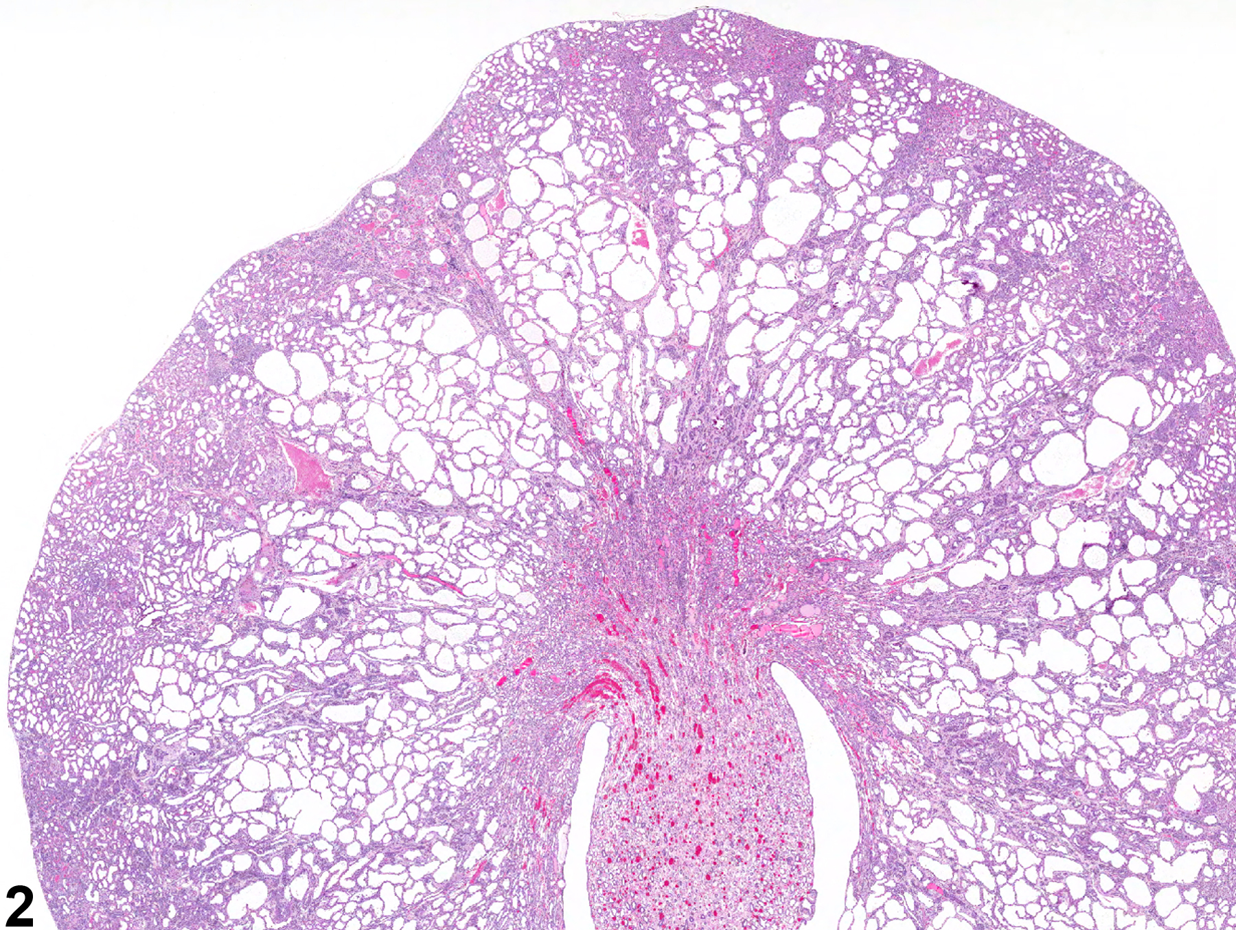
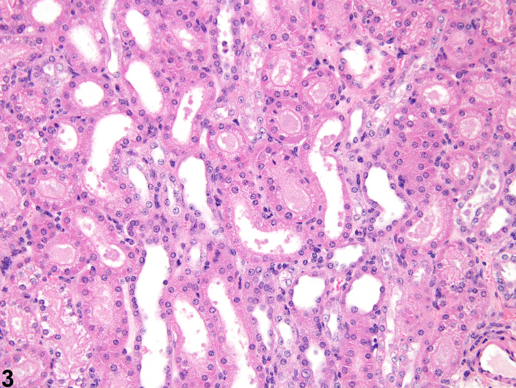
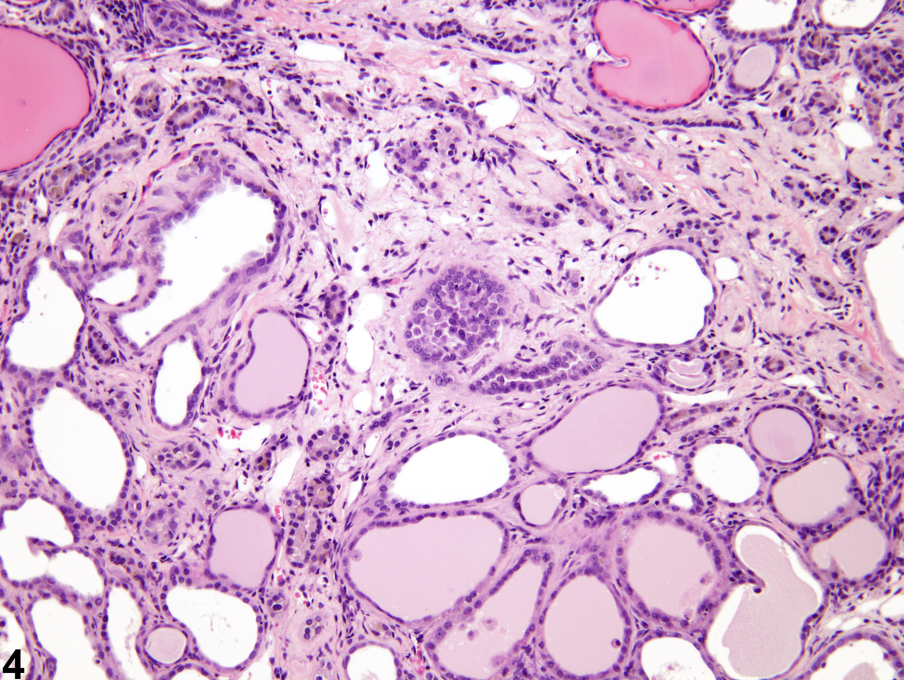
Renal tubule dilation may occur from xenobiotic administration, secondary mechanisms, or an unknown pathogenesis (see ) (Figure 2). Dilation may result from direct toxic injury to the tubule epithelium interfering with absorption and secretion (Figure 3). It may also occur secondary to renal ischemia or from prolonged diuresis related to drug administration. Secondary mechanisms of tubule dilation may result from lower urinary tract obstruction, the deposition of tubule crystals, interstitial inflammation and/or fibrosis, and chronic progressive nephropathy (Figure 4). A few dilated tubules may be regarded as normal histologic variation.
Greaves P. 2012. Histopathology of Preclinical Toxicity Studies, 4th ed. Elsevier, Amsterdam, 560.
Abstract: http://www.sciencedirect.com/science/book/9780444538567Lameire N. 2005. The pathophysiology of acute renal failure. Crit Care Clinics 21:197-210.
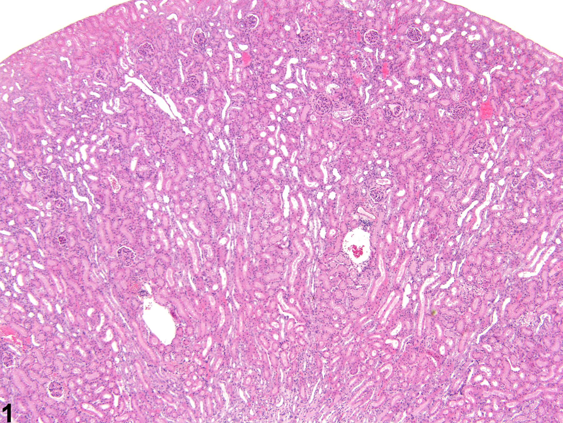
Kidney, Renal tubule - Dilation in a male B6C3F1 mouse from a chronic study. Dilated tubules are noted as tracts running through the cortex and outer medulla.
All Images

Kidney, Renal tubule - Dilation in a male B6C3F1 mouse from a chronic study. Dilated tubules are noted as tracts running through the cortex and outer medulla.

Kidney, Renal tubule - Dilation in a male F344/N rat from a chronic study. Tubule dilation is present throughout the outer stripe of the outer medulla, extending into the cortex.

Kidney, Renal tubule - Dilation in a male B6C3F1 mouse from a chronic study. Slight tubule dilation is associated with degeneration and necrosis.

Kidney, Renal tubule - Dilation in a male F344/N rat from a chronic study. Tubule dilation is associated with chronic progressive nephropathy.