

Urinary System
Kidney, Renal Tubule - Hypertrophy
Narrative
Ellison DH, Velazquez H, Wright FS. 1989. Adaptation of the distal tubule of the rat. Structural and functional effects of dietary salt intake and chronic diuretic infusion. J Clin Invest 83:113-126.
Full Text: http://www.ncbi.nlm.nih.gov/pmc/articles/PMC303651/Frazier KS, Seely JC, Hard GC, Betton G, Burnett R, Nakatsuji S, Nishikawa A, Durchfeld-Meyer B, Bube A. 2012. Proliferative and non-proliferative lesions in the rat and mouse urinary system. Toxicol Pathol 40:14S-86S.
Abstract: http://www.ncbi.nlm.nih.gov/pubmed/22637735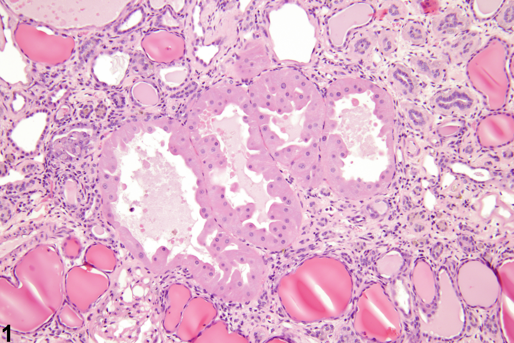
Kidney, Renal tubule - Hypertrophy in a male F344/N rat from a chronic study. These hypertrophied tubular epithelial cells with an increased amount of amorphous, eosinophilic cytoplasm and small, round, dense nuclei are associated with chronic progressive nephropathy.
All Images

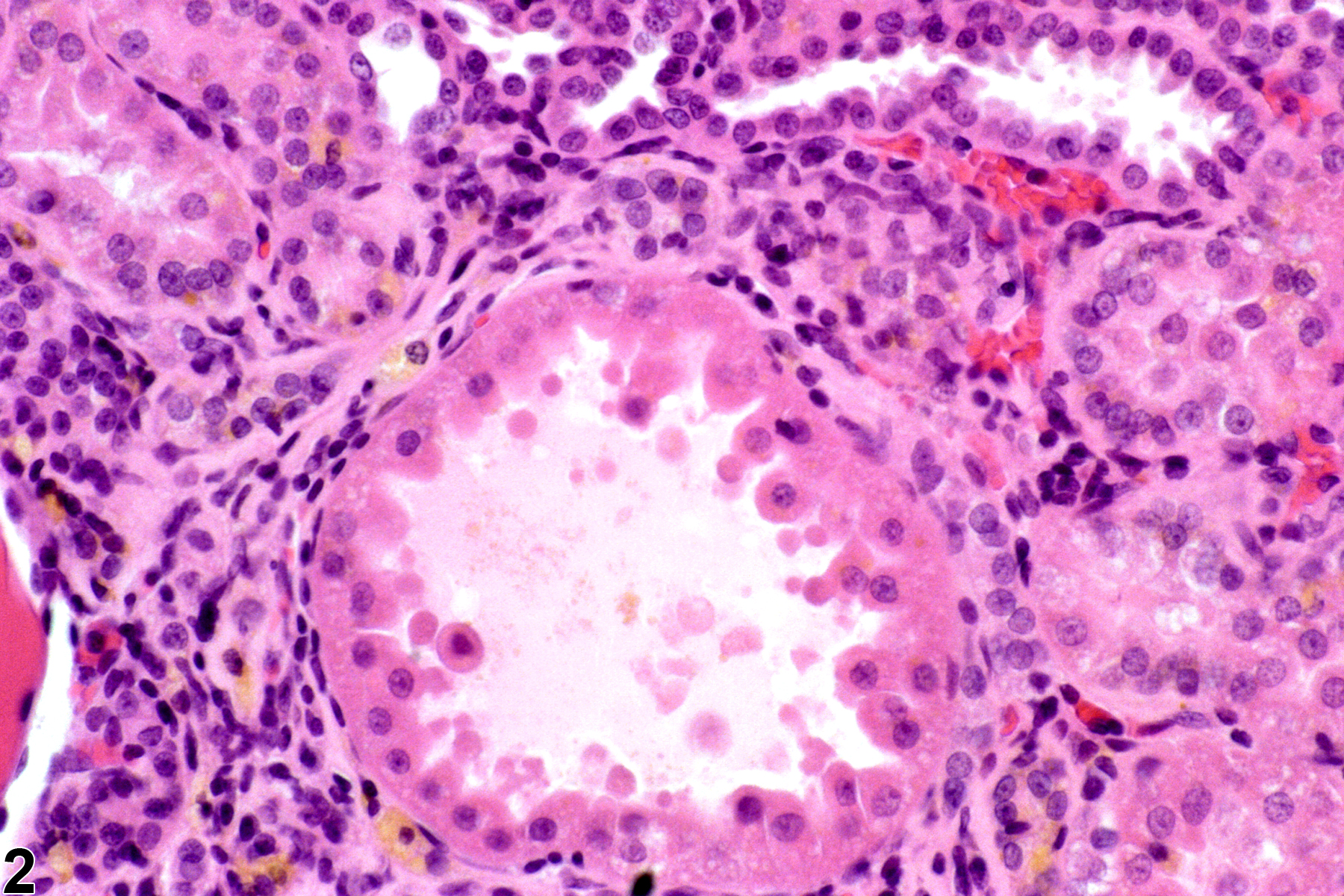
Kidney, Renal tubule - Hypertrophy in a male F344/N rat from a chronic study. These hypertrophied tubular epithelial cells with an increased amount of amorphous, eosinophilic cytoplasm and small, round, dense nuclei are associated with chronic progressive nephropathy.

Kidney, Renal tubule - Hypertrophy in a male F344/N rat. This is a slightly higher magnification example of a hypertrophied tubule.