

Urinary System
Kidney, Renal Tubule - Vacuolation, Cytoplasmic
Narrative
Bendele A, Seely J, Richey C, Sennello G, Shopp G. 1998. Short communication: Renal tubular vacuolation in animals treated with polyethylene-glycol-conjugated proteins. Toxicol Sci 42:152-157.
Abstract: http://www.ncbi.nlm.nih.gov/pubmed/9579027Johson RC, Dovey-Hartman BJ, Syed J, Leach MW, Frank DW, Sinha DP, Mirro EJ, Little JM, Halliwell WH. 1998. Vacuolation in renal tubular epithelium of Cd-1 mice: An incidental finding. Toxicol Pathol 26:789-792.
Full Text: http://tpx.sagepub.com/content/26/6/789.full.pdfKhan KNM, Alden CL. 2002. Kidney. In: Handbook of Toxicologic Pathology, 2nd ed (Haschek WG, Rousseaux CG, Wallig MA, eds). Academic Press, San Diego, 255-336.
Abstract: http://www.sciencedirect.com/science/book/9780123302151Monserrat AJ, Chandler AE. 1975. Effects of repeated injections of sucrose in the kidney: Histologic, cytochemical and functional studies in an animal model. Virchows Arch B 19:77-91.
Abstract: http://www.ncbi.nlm.nih.gov/pubmed/809924Nonoyama T, Fukuda R. 2008. Drug-induced phospholipidosis - pathological aspects and its prediction. J Toxicol Pathol 21:9-24.
Abstract: https://www.jstage.jst.go.jp/article/tox/21/1/21_1_9/_articleRees JA, Old SI, Rowlands PC. 1997. An ultrastructural, histochemistry, and light microscopy study of the early development of renal proximal tubular vacuolation after a single administration of the contrast enhancement medium "Iotrolan." Toxicol Pathol 25:158-164.
Abstract: http://www.ncbi.nlm.nih.gov/pubmed/9125774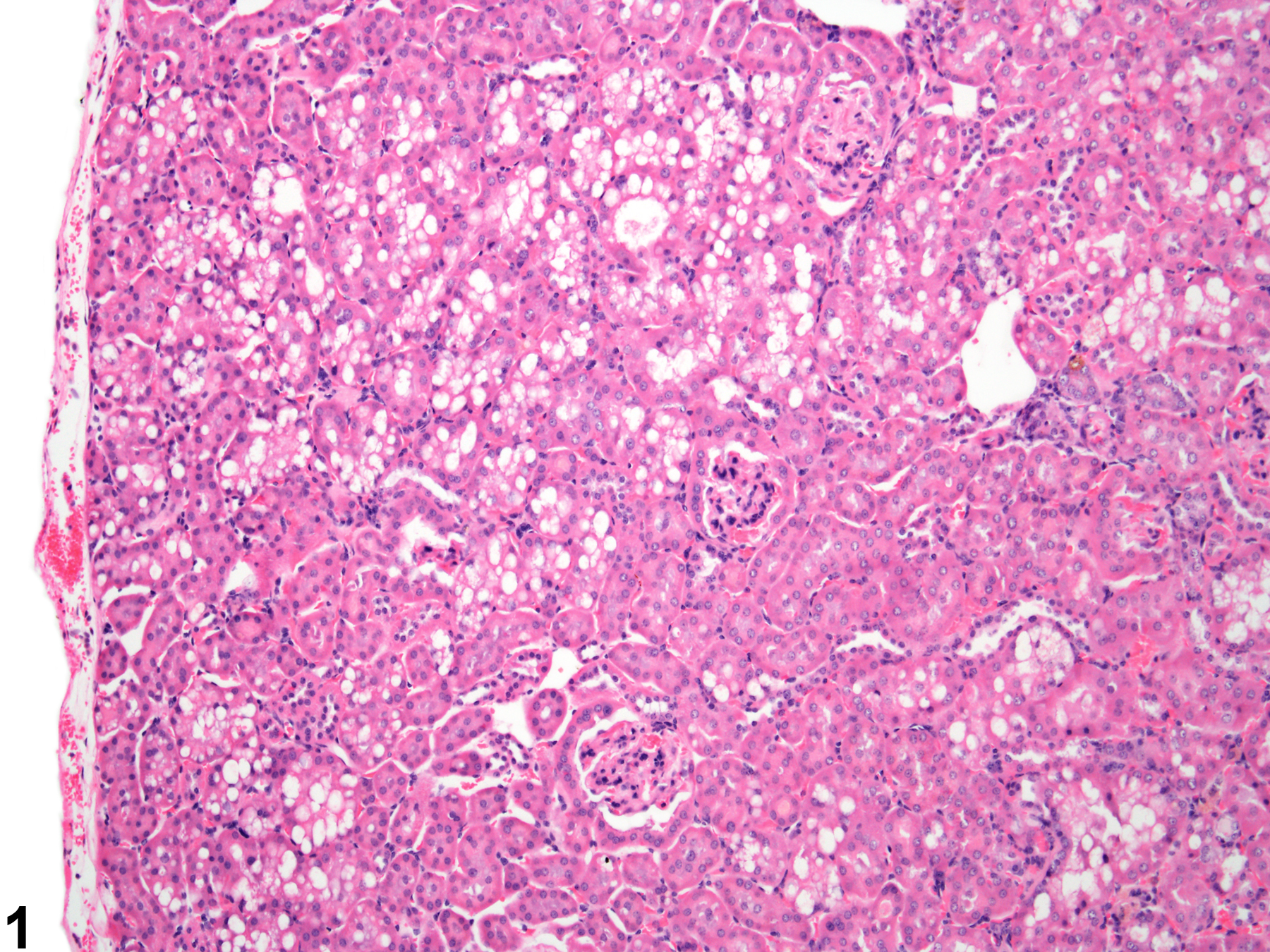
Kidney, Renal tubule - Vacuolation, Cytoplasmic in a treated male B6C3F1 mouse from a chronic study. Numerous clear vacuoles are present in the cytoplasm of renal tubule epithelial cells.
All Images

Kidney, Renal tubule - Vacuolation, Cytoplasmic in a treated male B6C3F1 mouse from a chronic study. Numerous clear vacuoles are present in the cytoplasm of renal tubule epithelial cells.
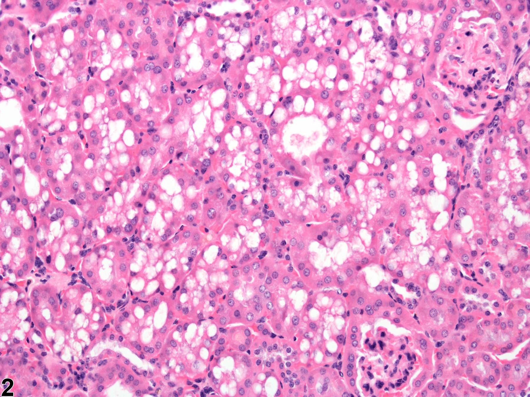
Kidney, Renal tubule - Vacuolation, Cytoplasmic in a treated male B6C3F1 mouse from a chronic study (higher magnification of Figure 1). There are variably sized, mostly large vacuoles in the renal tubule epithelial cells.