U.S. flag

An official website of the United States government

Dot gov

The .gov means it's official.
Federal government websites often end in .gov or .mil. Before sharing sensitive information, make sure you're on a federal government site.

Https

The site is secure.
The https:// ensures that you are connecting to the official website and that any information you provide is encrypted and transmitted securely.

Skip to Main Navigation
Skip to Page Content
Skip to Atlas Navigation

Urinary Bladder - Inflammation

Image of inflammation in the urinary bladder from a male F344/N rat in a chronic study
Chronic-active inflammation involving the urothelium and subepithelial layers from a male F344/N rat in a chronic study.
Figure 1 of 2
Image of inflammation in the urinary bladder from a male F344/N rat in a chronic study
Acute inflammation within the urothelium from a male F344/N rat in a chronic study.
Figure 2 of 2
next arrow

comment:

Inflammation is one of the most frequently diagnosed lesions of the urinary bladder. Inflammation may be acute, suppurative, chronic, chronic-active, or granulomatous, depending on the predominant cell type or cell response involved. In acute inflammation, the predominant infiltrating cell is the neutrophil, though fewer macrophages and lymphocytes may also be present. There may also be evidence of edema and hyperemia. The neutrophil is also the predominant cell type in suppurative inflammation; however, the neutrophils are aggregated, and many of them are degenerative (suppurative exudate). Lymphocytes predominate in chronic inflammation; lymphocytes also predominate in chronic-active inflammation, but there are also a significant number of neutrophils—both lesions contain macrophages. Diagnosis of granulomatous inflammation, another form of chronic inflammation, requires the presence of a significant number of aggregated, large, activated macrophages, epithelioid macrophages, or multinucleated giant cells.

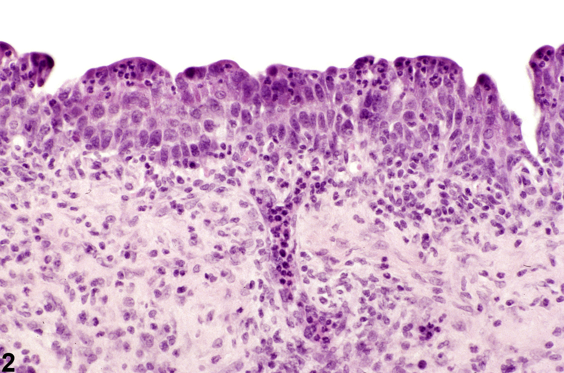
Inflammatory cells can be seen anywhere in the bladder, including the lumen, mucosa, submucosa, and muscularis ( Figure 1image opens in a pop-up window ). Hemorrhage, necrosis, urothelial hyperplasia, and fibrosis may be evident, depending on the lesion ( Figure 2image opens in a pop-up window ). In most instances, inflammation arises from bacterial infections or the presence of calculi. Inflammation may also be a direct consequence of chemical administration.

recommendation:

Inflammation should be diagnosed according to the cell response and the location within the bladder and should be given a severity grade. Hemorrhage, necrosis, urothelial hyperplasia, and fibrosis may be diagnosed separately if they constitute a significant component of the lesion.

references:

Frith CH. 1979. Morphologic classification of inflammatory, nonspecific, and proliferative lesions of the urinary bladder of mice. Invest Urol 16:435-444.
Abstract: http://www.ncbi.nlm.nih.gov/pubmed/571862

Gaillard ET. 1999. Ureter, urinary bladder and urethra. In: Pathology of the Mouse: Reference and Atlas (Maronpot RR, Boorman GA Gaul BW, eds). Cache River Press, Vienna, IL, 235-258.
Abstract: http://www.cacheriverpress.com/books/pathmouse.htm

Jokinen MP. 1990. Urinary bladder, ureter, and urethra. In: Pathology of the Fischer Rat: Reference and Atlas (Boorman GA, Eustis SL, Elwell MR, Montgomery CA, MacKenzie WF, eds). Academic Press, San Diego, 109-126.
Abstract: http://www.ncbi.nlm.nih.gov/nlmcatalog/9002563
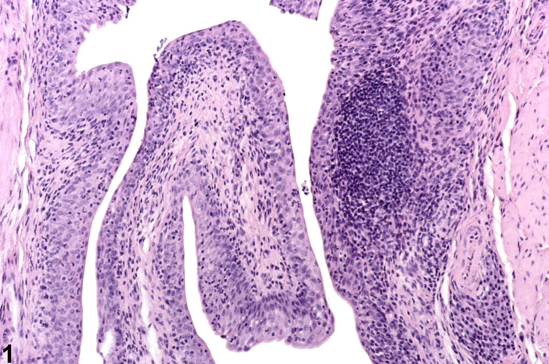