U.S. flag

An official website of the United States government

Dot gov

The .gov means it's official.
Federal government websites often end in .gov or .mil. Before sharing sensitive information, make sure you're on a federal government site.

Https

The site is secure.
The https:// ensures that you are connecting to the official website and that any information you provide is encrypted and transmitted securely.

Skip to Main Navigation
Skip to Page Content
Skip to Atlas Navigation

Urinary Bladder - Metaplasia

Image of metaplasia in the urinary bladder from a female F344/N rat in a chronic study
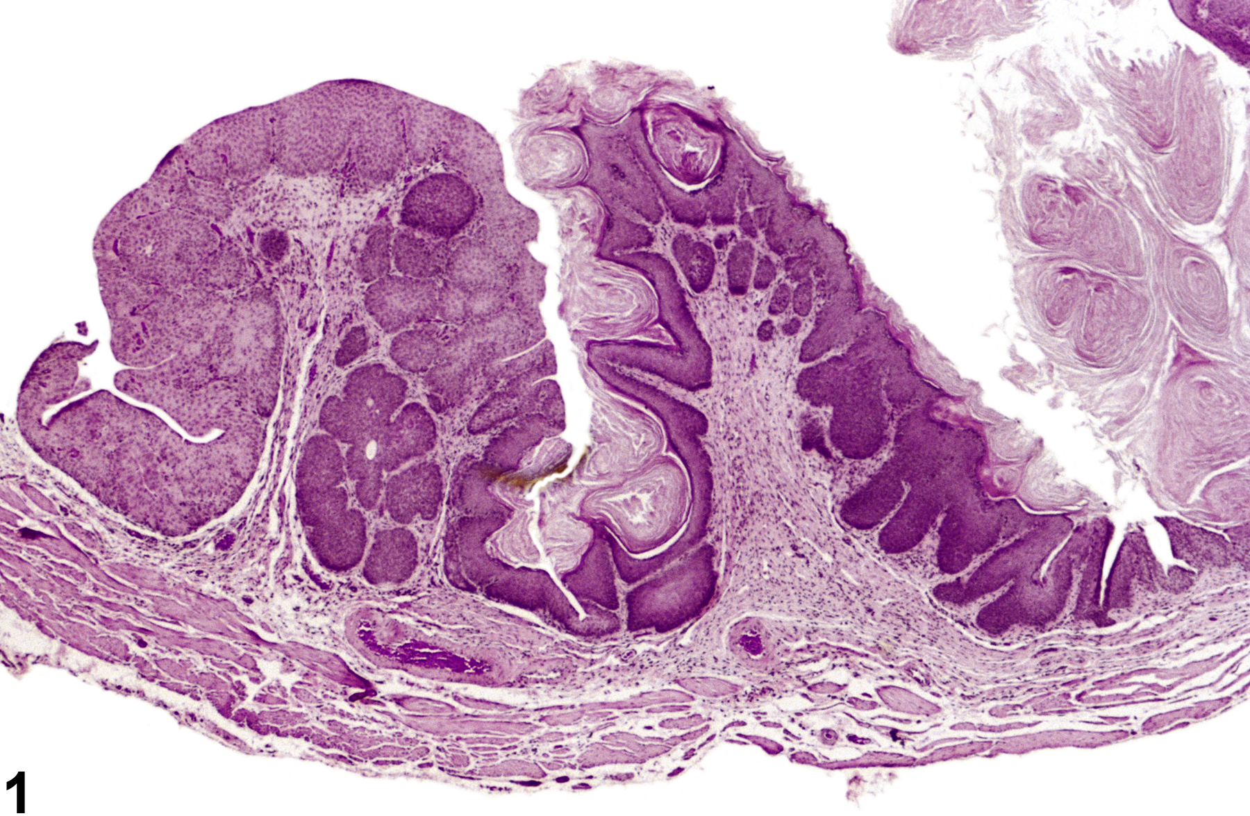
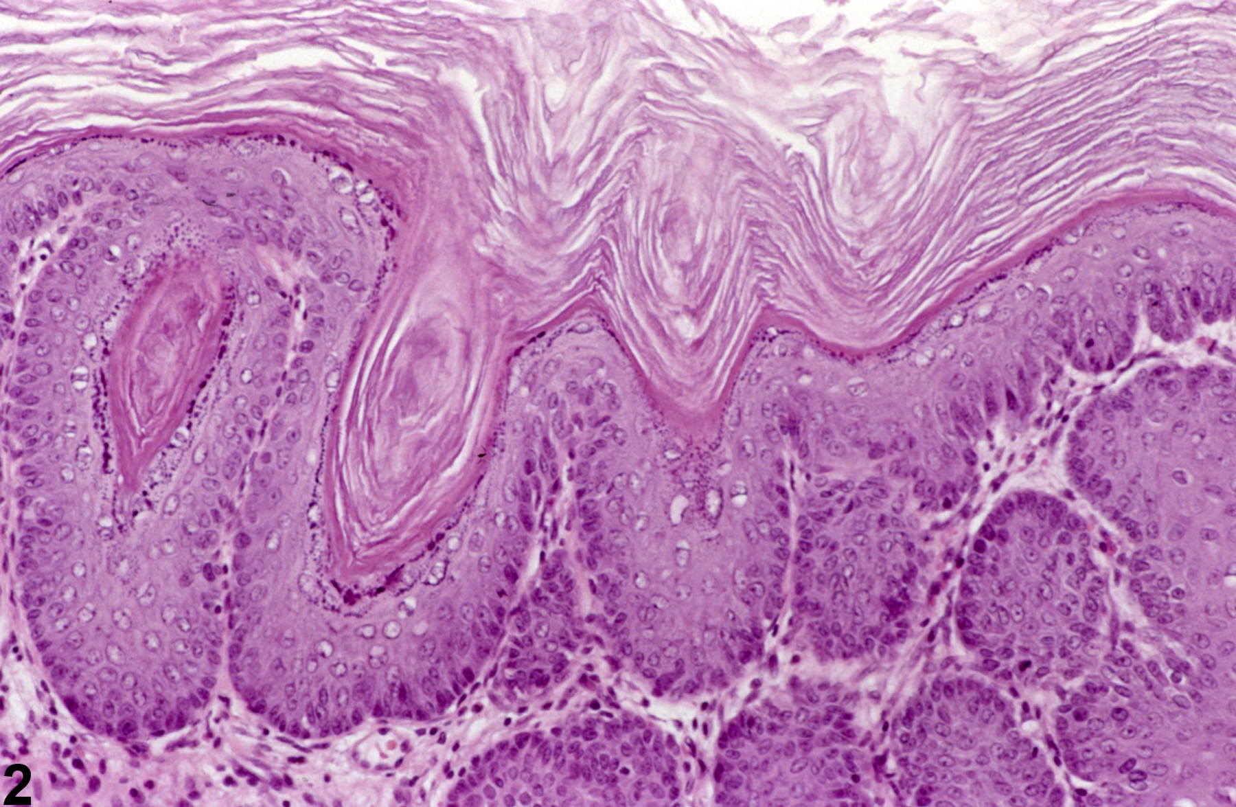
Diffuse squamous metaplasia and keratinization of hyperplastic urothelium from a female F344/N rat in a subchronic interim study.
Figure 1 of 4
Image of metaplasia in the urinary bladder from a female F344/N rat in a chronic study
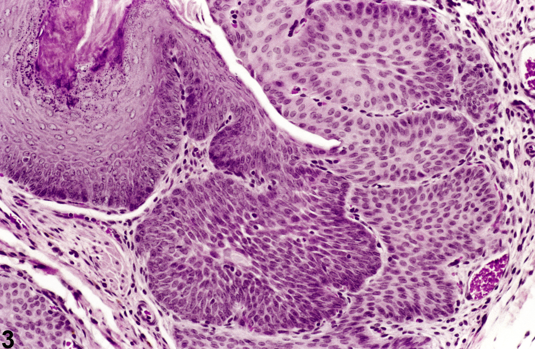
Squamous metaplasia and keratinization from a female F344/N rat in a 40-week interim study.
Figure 2 of 4
Image of metaplasia in the urinary bladder from a female F344/N rat in a chronic study
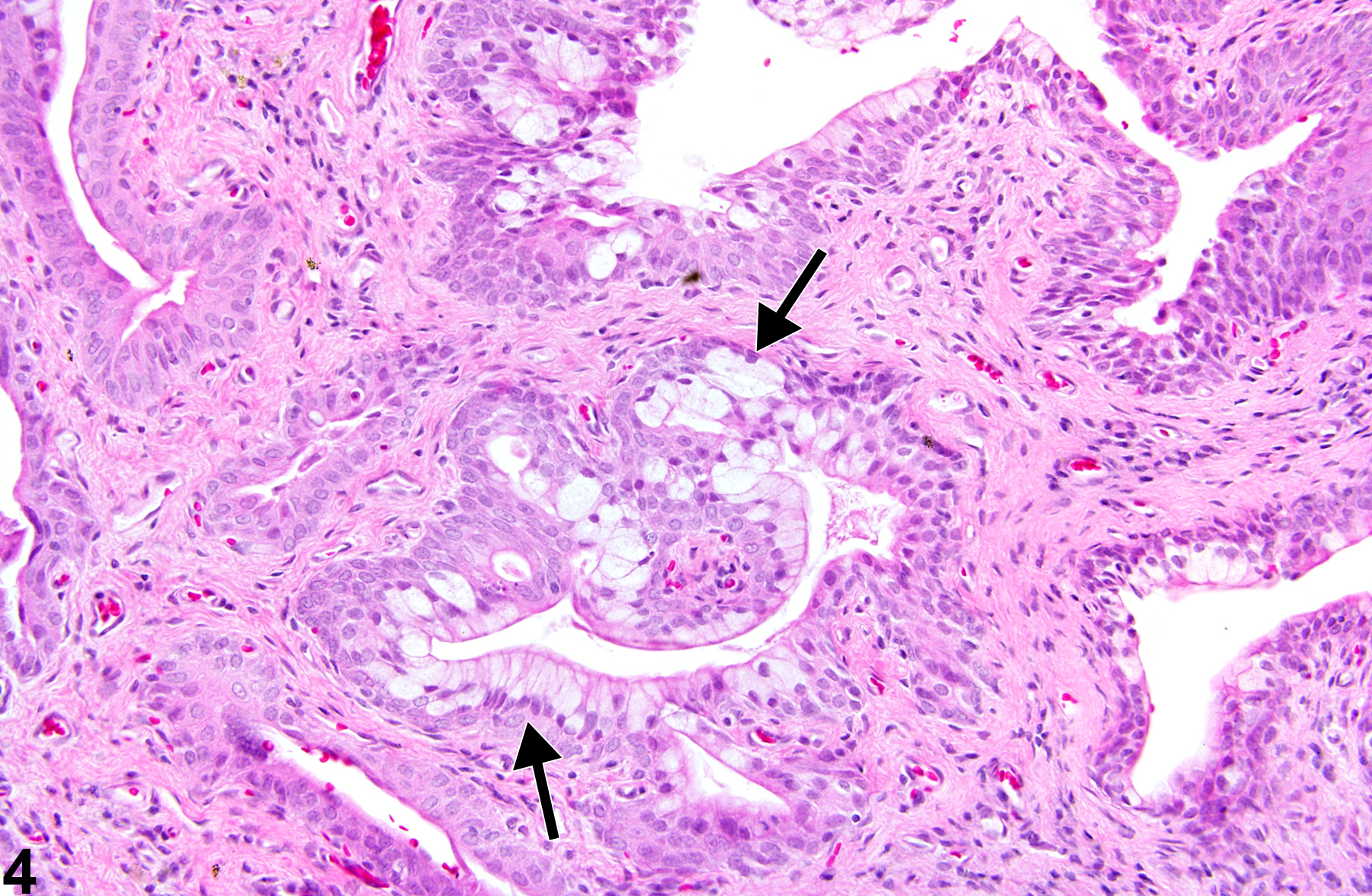
Squamous metaplasia with and without keratinization, from a female F344/N rat in a subchronic interim study.
Figure 3 of 4
Image of metaplasia in the urinary bladder from a female F344/N rat in a chronic study
Mucinous metaplasia (arrows) characterized by mucinous cells in areas of urothelial hyperplasia, from a female F344/N rat in a chronic study.
Figure 4 of 4
next arrow

comment:

Metaplasia of the urothelium reflects the potential of the urothelium to undergo a metaplastic change under a variety of conditions. Squamous metaplasia, with or without keratinization, is the most common variant observed ( Figure 1image opens in a pop-up window , Figure 2image opens in a pop-up window and , Figure 3image opens in a pop-up window ). Mucinous, glandular, or mixtures of metaplastic lesions are less commonly observed ( Figure 4image opens in a pop-up window ).

recommendation:

Urothelial metaplasia and, in particular, the type of metaplasia should be diagnosed and given a severity grade when it becomes a major part of a lesion, for instance, in association with inflammation or hyperplasia. Small or focal lesions, when part of a primary lesion such as chronic inflammation and/or hyperplasia, should not be diagnosed separately.

references:

Gaillard ET. 1999. Ureter, urinary bladder and urethra. In: Pathology of the Mouse: Reference and Atlas (Maronpot RR, Boorman GA Gaul BW, eds). Cache River Press, Vienna, IL, 235-258.
Abstract: http://www.cacheriverpress.com/books/pathmouse.htm

Jokinen MP. 1990. Urinary bladder, ureter, and urethra. In: Pathology of the Fischer Rat: Reference and Atlas (Boorman GA, Eustis SL, Elwell MR, Montgomery CA, MacKenzie WF, eds). Academic Press, San Diego, 109-126.
Abstract: http://www.ncbi.nlm.nih.gov/nlmcatalog/9002563