U.S. flag

An official website of the United States government

Dot gov

The .gov means it's official.
Federal government websites often end in .gov or .mil. Before sharing sensitive information, make sure you're on a federal government site.

Https

The site is secure.
The https:// ensures that you are connecting to the official website and that any information you provide is encrypted and transmitted securely.

Skip to Main Navigation
Skip to Page Content
Skip to Atlas Navigation

Urinary Bladder, Urothelium - Hyperplasia

Image of hyperplasia in the urinary bladder from a male F344/N rat in a chronic study
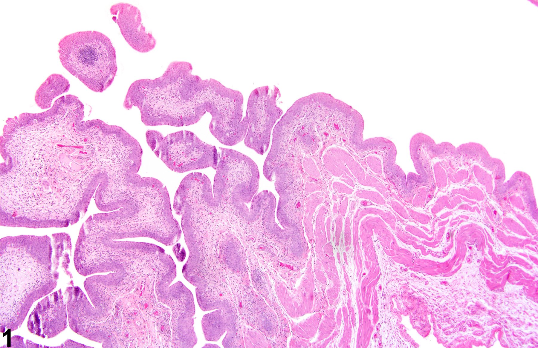
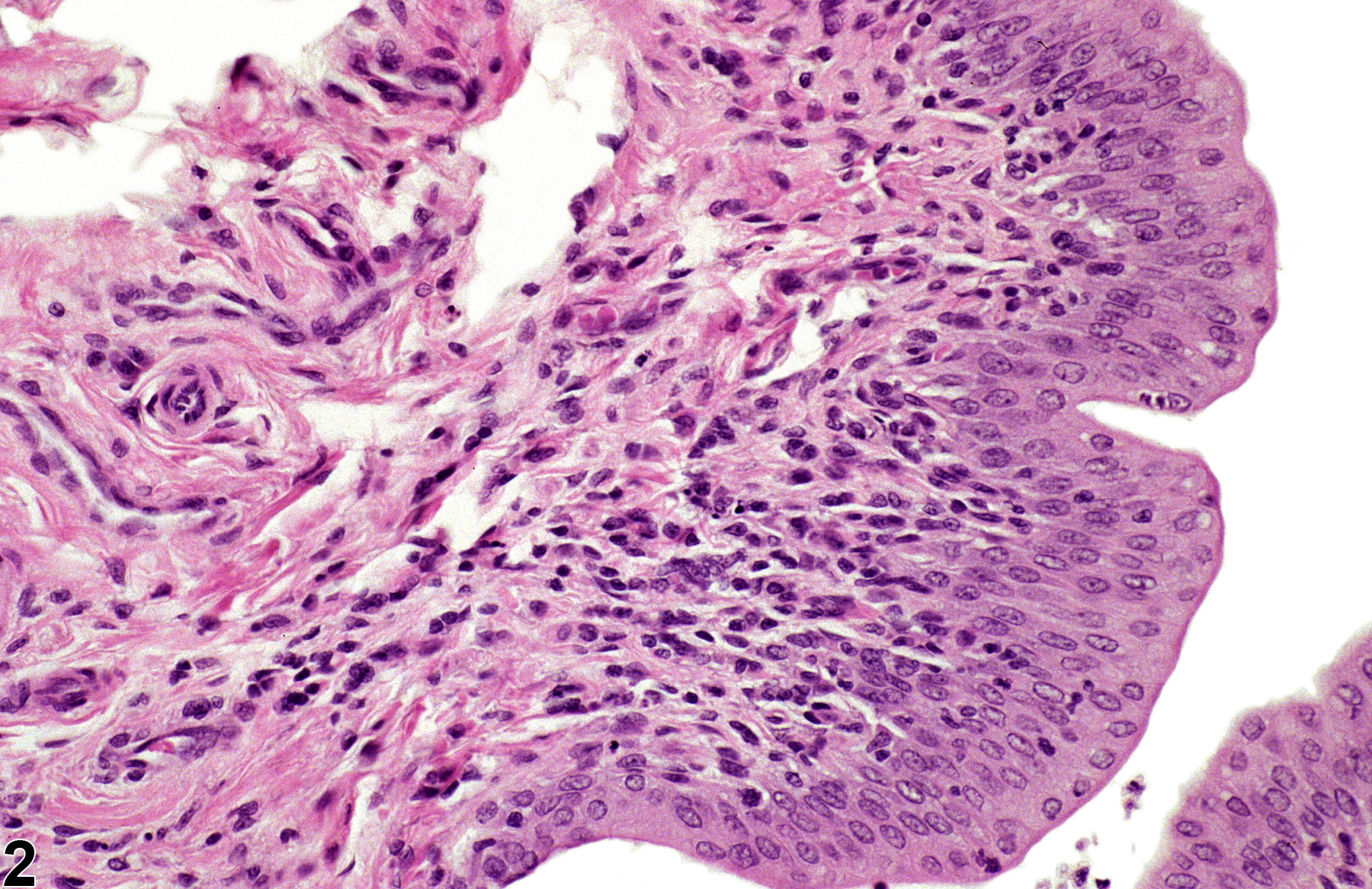
Urothelial hyperplasia (simple) from a male F344/N rat in a chronic study.
Figure 1 of 4
Image of hyperplasia in the urinary bladder from a female Harlan Sprague-Dawley rat in a chronic study
Urothelial hyperplasia (simple) with minimal inflammatory infiltration of epithelium and submucosa, from a female Harlan Sprague-Dawley rat in a chronic study.
Figure 2 of 4
Image of hyperplasia in the urinary bladder from a male F344/N rat in a chronic study
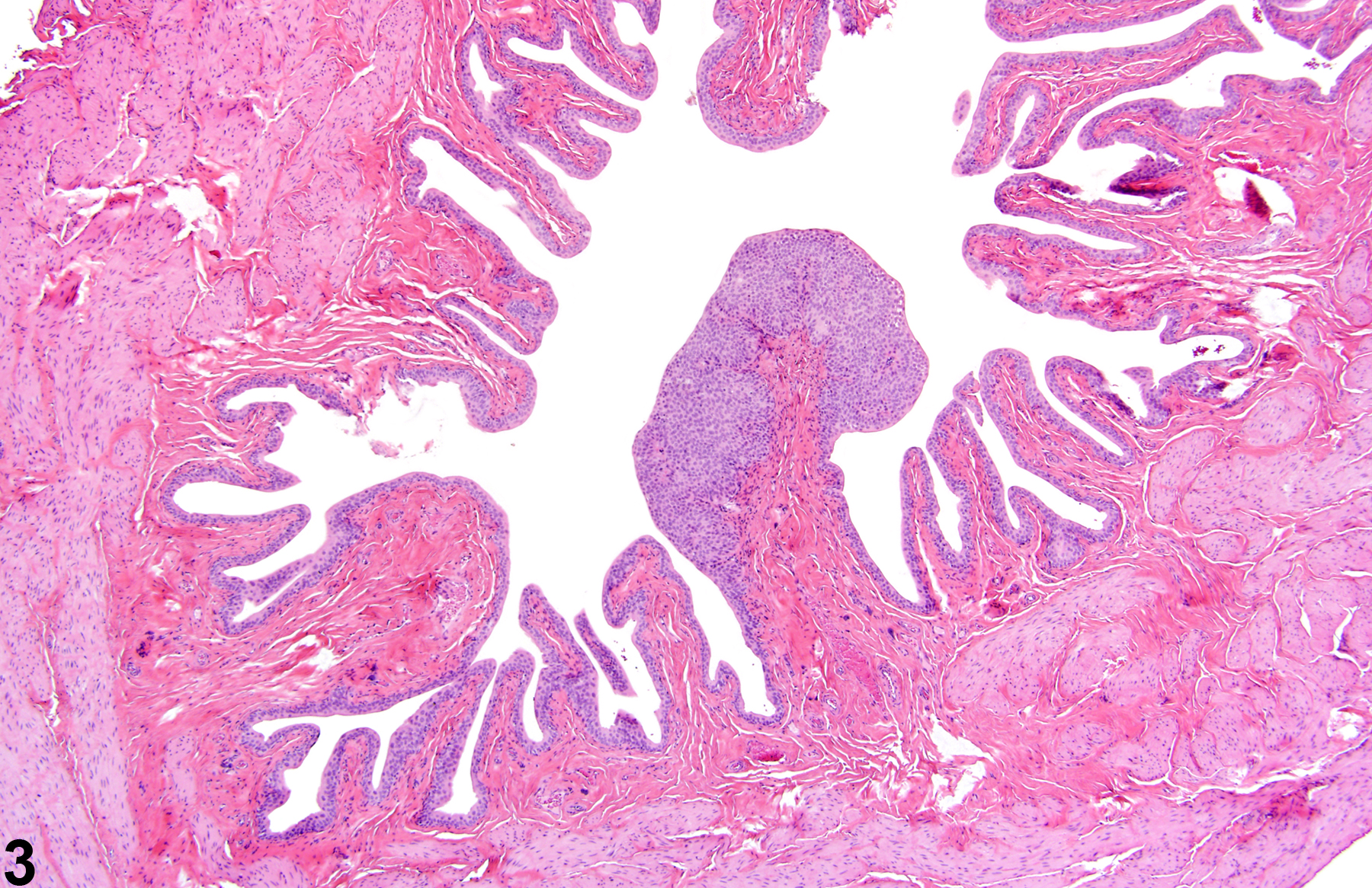
Urothelial hyperplasia (nodular) from a male F344/N rat in a chronic study.
Figure 3 of 4
Image of hyperplasia in the urinary bladder from a male TG/RASH2/CB6F1 mouse
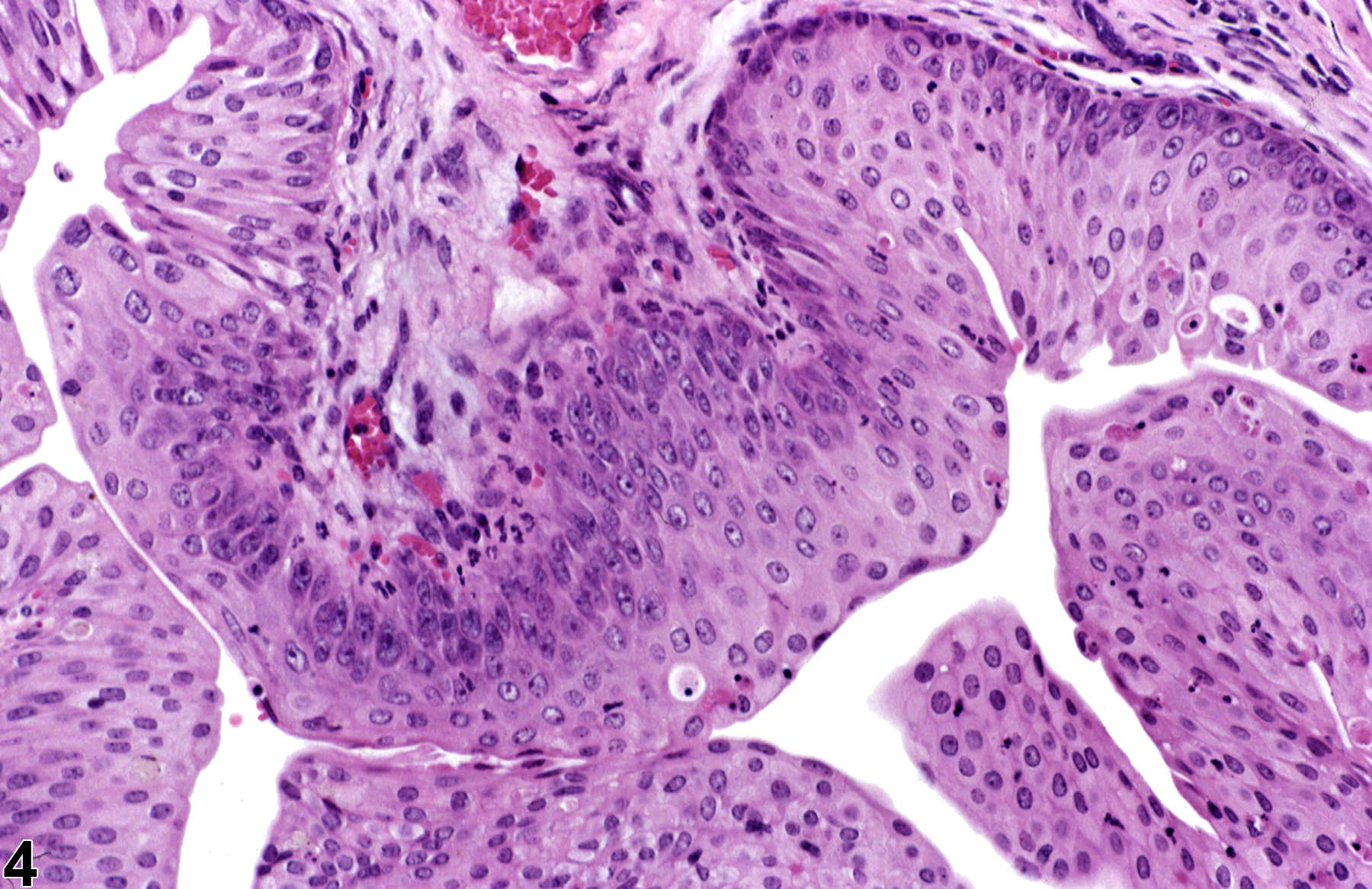
Urothelial hyperplasia with little to no inflammation, from a male TG/RASH2/CB6F1 mouse.
Figure 4 of 4
next arrow

comment:

Urothelial hyperplasia, defined as an increased number of epithelial cells, is commonly observed with inflammation, with the presence of calculi, or as a response to chemical administration. In general, hyperplasia can either be simple, nodular, or papillary ( Figure 1image opens in a pop-up window , Figure 2image opens in a pop-up window , Figure 3image opens in a pop-up window , and Figure 4image opens in a pop-up window ), which may not be important to diagnose for occasional and spontaneous cases. Most cases of hyperplasia are focal to diffuse. Hyperplastic cells may be normal in appearance or pleomorphic, or (rarely) may have atypia. Mitotic figures are variable. Metaplastic foci of squamous, glandular, or mixtures of cell metaplasia may be noted. Urothelial hyperplasia may be reversible but may also progress to neoplasia. Hyperplasia is less commonly observed in the mouse. Minimal hyperplasia is difficult to diagnose because of the variability of the bladder appearance during fixation.

recommendation:

Usually a guideline of an increase above the normal 3- to 4-cell-thick urothelium constitutes hyperplasia. However, sectioning also poses a difficulty in determining minimal or borderline hyperplasias because of folding of the bladder lining in nondistended states. Care should be taken to not confuse tangential sectioning as hyperplasia. Hyperplasia should be diagnosed and given a severity grade. Hyperplastic modifiers such as “simple,” “nodular,” or “papillary” associated with chemical exposure should be discussed in the narrative report.

references:

Cohen SM. 1989. Toxic and nontoxic changes induced in the urothelium by xenobiotics. Toxicol Appl Pharmacol 101:484-498.
Abstract: http://www.ncbi.nlm.nih.gov/pubmed/2603162

Cohen SM. 2002. Comparative pathology of proliferative lesions of the urinary bladder. Toxicol Pathol 30:663-671.
Abstract: http://www.ncbi.nlm.nih.gov/pubmed/12512866

Cohen SM, Cano M, Anderson T, Garland EM. 1996. Extensive handling of rats leads to mild urinary bladder hyperplasia. Toxicol Pathol 24:251-257.
Abstract: http://www.ncbi.nlm.nih.gov/pubmed/8992616

Gaillard ET. 1999. Ureter, urinary bladder and urethra. In: Pathology of the Mouse: Reference and Atlas (Maronpot RR, Boorman GA Gaul BW, eds). Cache River Press, Vienna, IL, 235-258.
Abstract: http://www.cacheriverpress.com/books/pathmouse.htm

Greaves P. 2007. Histopathology of Preclinical Studies, 3rd ed. Elsevier, Amsterdam, 591-592.
Abstract: http://www.sciencedirect.com/science/book/9780444538567

Jokinen MP. 1990. Urinary bladder, ureter, and urethra. In: Pathology of the Fischer Rat: Reference and Atlas (Boorman GA, Eustis SL, Elwell MR, Montgomery CA, MacKenzie WF, eds). Academic Press, San Diego, 109-126.
Abstract: http://www.ncbi.nlm.nih.gov/nlmcatalog/9002563

Kunze E. 1998. Hyperplasia, urinary bladder, rat. In: Monographs on Pathology of Laboratory Animals: Urinary System, 2nd ed (Jones TC, Hard GC, Mohr U, eds). Springer, Berlin, 331-368.
Abstract: http://www.ilsi.org/publications/urinarysystem.pdf

Shinohara Y, Frith CH. 1981. Comparison of experimental and spontaneous bladder urothelial hyperplasias occurring in BALB/c mice. Invest Urol 18:233-238.
Abstract: http://www.ncbi.nlm.nih.gov/pubmed/7451083