Integumentary System
Mammary Gland - Dilation
Narrative
Benign mammary gland dilation is a common age-related change in animals. As animals age, in the process of involution, the composition of the mammary gland changes from mostly glandular to mostly fatty, which can result in blockage of a duct, intraluminal accumulations, and dilation. Additionally, the age-related changes of mammary gland dilation may be associated with mammary gland hyperplasia and metaplasia. In younger animals treated with xenobiotics, duct dilation suggests perturbation of the hypothalamic-pituitary-gonadal axis.
Mammary gland dilation can affect ducts, alveoli, or both. It is often a diffuse change characterized by distention of collecting (lactiferous) ducts and alveoli beneath a nipple by intraluminal accumulations of amorphous, proteinaceous eosinophilic, secretory material, lipid, cell debris, and, sometimes, inflammatory cells. The lining epithelial cells of duct dilation are often vacuolated and can occur with or without epithelial hypertrophy or hyperplasia. Galactoceles are considered an extreme form of dilation and are characterized by focally dilated mammary gland ducts and alveoli that have become cystic and very large, lined by flattened epithelium, and filled with proteinaceous secretory fluid. Galactoceles may rupture and be associated with inflammation and fibrosis.
| Barsoum NJ, Gough AW, Sturgess JM, de la Iglesia FA. 1984. Morphologic features and incidence of spontaneous hyperplastic and neoplastic mammary gland lesions in Wistar rats. Toxicol Pathol 12(1):26-38. |
| Boorman GA, Wilson JT, Van Zwieten M, Eustis SL. 1990. Mammary gland. In: Boorman GA, Eustis SL, Elwell MR, Montgomery CA, Mackenzie WF (eds.). 2016. Pathology of the Fischer rat - reference and atlas. Academic Press pp. 295-313. |
| Burek JD. 1978. Pathology of aging rats. CRC Press pp. 163-167. |
| Yi ES, Bedoya AA, Lee H, Kim S, Housley RM, Aukerman SL, Tarpley JE, Starnes C, Yin S, Pigrce GF, Ulich TR. 1994. Keratinocyte growth factor causes cystic dilatation of the mammary Glands of mice. Am J Pathol 145(5):1015-1022. |
| Goodman DG, Ward JM, Squire RA, Chu KC, Linhart MS. 1979. Neoplastic and nonneoplastic lesions in aging F344 rats. Toxicol Appl Pharmacol 48(2):237-248. |
| Greaves P. 2007. Mammary gland. Histopathology of preclinical toxicity studies. Interpretation and relevance in drug safety evaluation, 3rd ed. Academic Press pp. 68-98. |
| Rehm S, Liebalt AG. Nonneoplastic and neoplastic lesions of the mammary gland. In: Mohr U, Dungworth DL, Ward J, Capen CC, Carlton WW, Sundberg JP (eds.). 1996. Pathobiology of the aging mouse, Vol. 2. International Life Sciences Institute Press pp. 381-398. |
| Russo J, Russo IH, Van Zwieten MJ, Rogers AE, Gusterson BA. Classification of neoplastic and nonneoplastic lesions of the rat mammary gland. In: Jones TC, Mohr U, Hunt RD (eds.). 1989. Integument and mammary glands (monographs on pathology of laboratory animals). International Life Sciences Institute pp. 275-304. |
| Seely JC, Boorman GA. Mammary gland and specialized sebaceous glands. In: Maronpot RR, Boorman GA, Gaul BW (eds.). 1999. Pathology of the mouse: reference and atlas. Cache River Press pp. 613-635. |
| Sourla A, Martel C, Labrie C, Labrie F. 1998. Almost exclusive androgenic action of dehydroepiandrosterone in the rat mammary gland. Endocrinology 139(2):753-64. Abstract: https://pubmed.ncbi.nlm.nih.gov/9449650 |
| Tucker MJ. 1997. The integumentary system and mammary glands. Diseases of the Wistar rat. CRC Press pp. 23-36. |
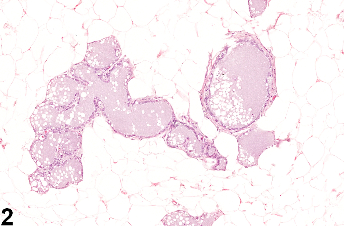
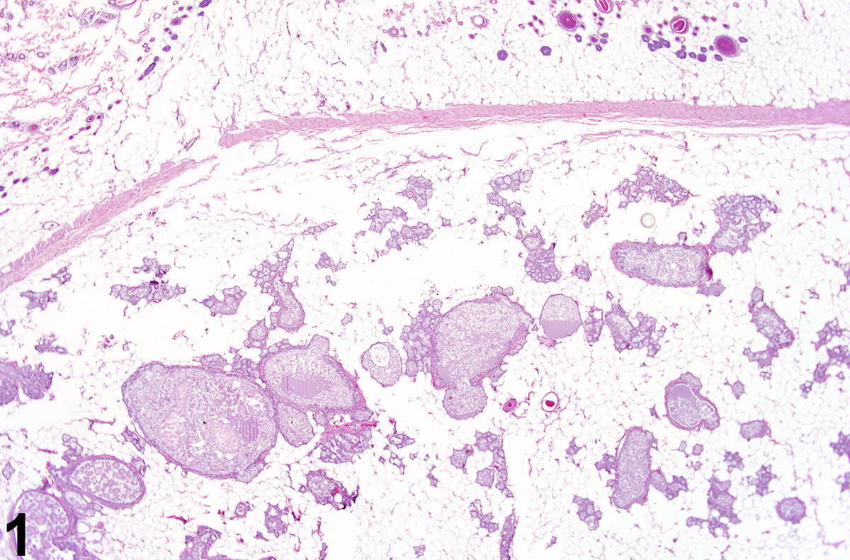 Mammary gland - Dilation in a female F344/N rat from a chronic study. There are scattered ducts and alveoli distended by intraluminal accumulations of amorphous, secretory material.
All Images