

Integumentary System
Mammary Gland - Hyperplasia, Atypical
Narrative
Al-Dhaheri WS, Hassouma I, Al-Salam S, Karam SM. 2008. Characterization of breast cancer progression in the rat. Ann N Y Acad Sci 1138:121-131.
Abstract: http://www.ncbi.nlm.nih.gov/pubmed/18837892Barsoum NJ, Gough AW, Sturgess JM, de la Iglesia FA. 1984. Morphologic features and incidence of spontaneous hyperplastic and neoplastic mammary gland lesions in Wistar rats. Toxicol Pathol 12(1):26-38.
Full Text: http://tpx.sagepub.com/content/12/1/26.full.pdfBoorman GA, Wilson JT, Van Zwieten M, Eustis SL. 1990. Mammary gland. In: Boorman GA, Eustis SL, Elwell MR, Montgomery CA, Mackenzie WF (eds.). 2016. Pathology of the Fischer rat - reference and atlas. Academic Press pp. 295-313.
Burek JD. 1978. Pathology of aging rats. CRC Press pp. 163-167.
Goodman DG, Ward JM, Squire RA, Chu KC, Linhart MS. 1979. Neoplastic and nonneoplastic lesions in aging F344 rats. Toxicol Appl Pharmacol 48(2):237-248.
Abstract: http://www.sciencedirect.com/science/article/pii/0041008X79900292Greaves P. 2007. Mammary gland. Histopathology of preclinical toxicity studies. Interpretation and relevance in drug safety evaluation, 3rd ed. Academic Press pp. 68-98.
Harvell DME, Strecker TE, Tochacek M, Xie B, Pennington KL, McComb RD, Roy SK, Shull JD. 2000. Rat strain-specific actions of 17β-estradiol in the mammary gland: correlation between estrogen-induced lobuloalveolar hyperplasia and susceptibility to estrogen-induced mammary cancers. Proc Natl Acad Sci U S A 97(6):2779-2784.
Abstract: http://www.ncbi.nlm.nih.gov/pmc/articles/PMC16006Latendresse JR, Bucci TJ, Olson G, Mellick P, Weis CC, Thorn B, Newbold RR, Delclos KB. 2009. Genistein and ethinyl estradiol dietary exposure in multigenerational and chronic studies induce similar proliferative lesions in mammary gland of male Sprague-Dawley rats. Reprod Toxicol 28(3):342-53.
Abstract: http://www.ncbi.nlm.nih.gov/pubmed/19383540Masso-Welch PA, Darcy KM, Stangle-Castor NC, Ip MM. 2000. A developmental atlas of rat mammary gland histology. J Mammary Gland Biol Neoplasia 5(2):165-85.
Abstract: http://www.ncbi.nlm.nih.gov/pubmed/11149571McMartin DN, Sahota PS, Gunson DE, Hsu HH, Spaet RH. 1992. Neoplasms and related proliferative lesions in control Sprague-Dawley rats from carcinogenicity studies. Historical data and diagnostic considerations. Toxicol Pathol 20(2):212-25.
Abstract: http://www.ncbi.nlm.nih.gov/pubmed/1475582National Toxicology Program Abstract for TR-485. 1999. Toxicology and carcinogenesis studies of oxymetholone (CAS No. 434 -07 -1) in F344/N rats and toxicology studies of oxymetholone in B6C3F1 mice (Gavage Studies).
Abstract: http://ntp.niehs.nih.gov/go/9771Rehm S, Liebalt AG. Nonneoplastic and neoplastic lesions of the mammary gland. In: Mohr U, Dungworth DL, Ward J, Capen CC, Carlton WW, Sundberg JP (eds.). 1996. Pathobiology of the aging mouse, Vol. 2. International Life Sciences Institute Press pp. 381-398.
Schedin P, Mitrenga T, Kaeck M. 2000. Estrous cycle regulation of mammary epithelial cell proliferation, differentiation, and death in the Sprague-Dawley rat: a model for investigating the role of estrous cycling in mammary carcinogenesis. J Mammary Gland Biol Neoplasia 5(2):211-25.
Abstract: http://www.ncbi.nlm.nih.gov/pubmed/11149574Seely JC, Boorman GA. Mammary gland and specialized sebaceous glands. In: Maronpot RR, Boorman GA, Gaul BW (eds.). 1999. Pathology of the mouse: reference and atlas. Cache River Press pp. 613-635.
Van Zwieten MJ, HogenEsch H, Majka JA, Boorman GA. Nonneoplastic and neoplastic lesions of the mammary gland. In: Mohr U, Dungworth DL, Capen CC (eds.). 1994. Pathobiology of the aging rat, Vol. 2. International Life Sciences Press pp. 459-474.
Harvell DME, Strecker TE, Tochacek M, Xie B, Pennington KL, McComb RD, Roy SK, Shull JD. 2000. Rat strain-specific actions of 17β-estradiol in the mammary gland: correlation between estrogen-induced lobuloalveolar hyperplasia and susceptibility to estrogen-induced mammary cancers. Proc Natl Acad Sci U S A 97(6):2779-2784.
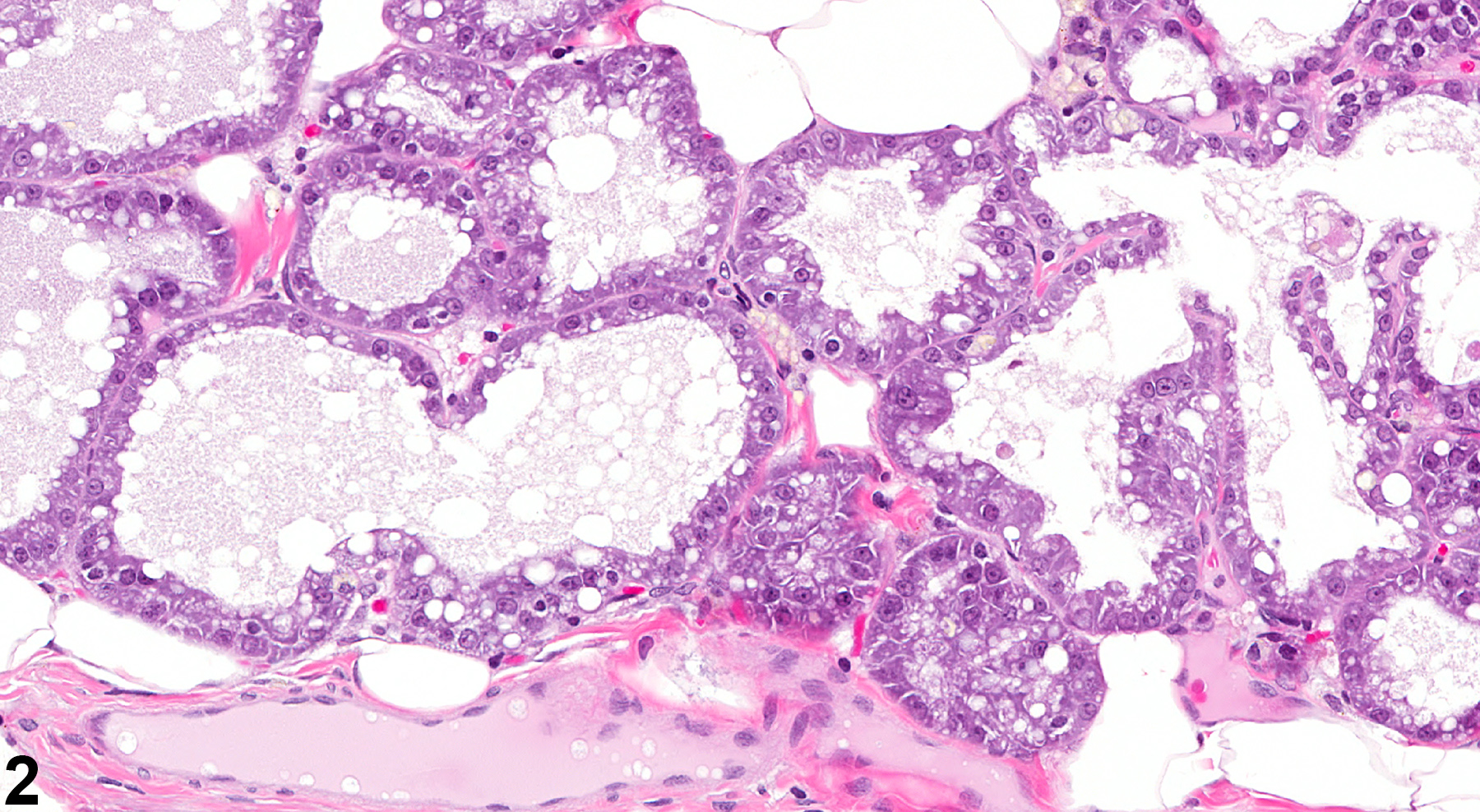
Abstract: http://www.ncbi.nlm.nih.gov/pmc/articles/PMC16006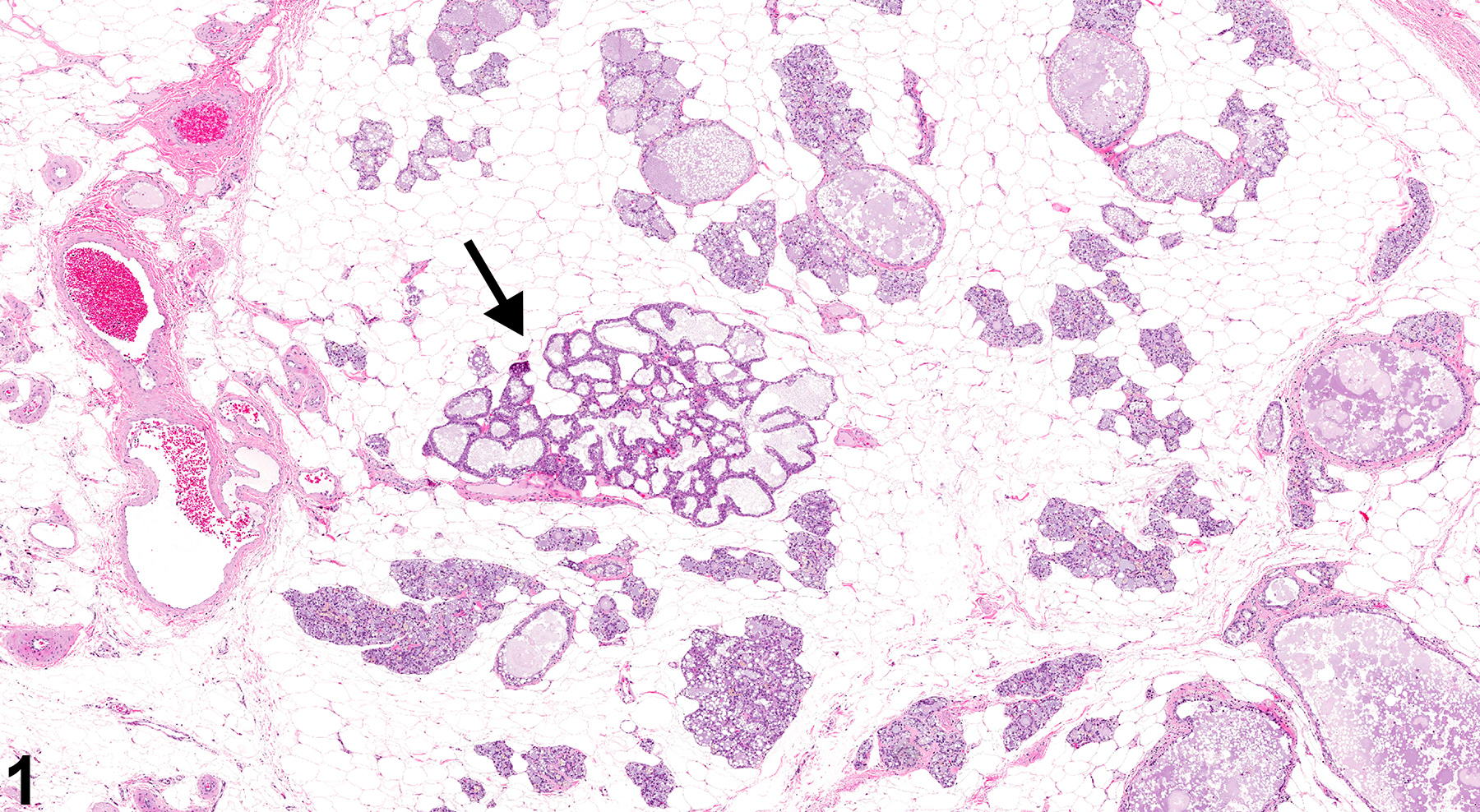 Mammary gland, Alveolus - Atypical Hyperplasia, in a female F344/N rat from a chronic study. There is a focal area of Atypical Hyperplasia (arrow) in the mammary gland.
All Images

