U.S. flag

An official website of the United States government

Dot gov

The .gov means it's official.
Federal government websites often end in .gov or .mil. Before sharing sensitive information, make sure you're on a federal government site.

Https

The site is secure.
The https:// ensures that you are connecting to the official website and that any information you provide is encrypted and transmitted securely.

Skip to Main Navigation
Skip to Page Content
Skip to Atlas Navigation

Esophagus - Inflammation

Image of inflammation in the esophagus from a female F344/N rat in a subchronic study
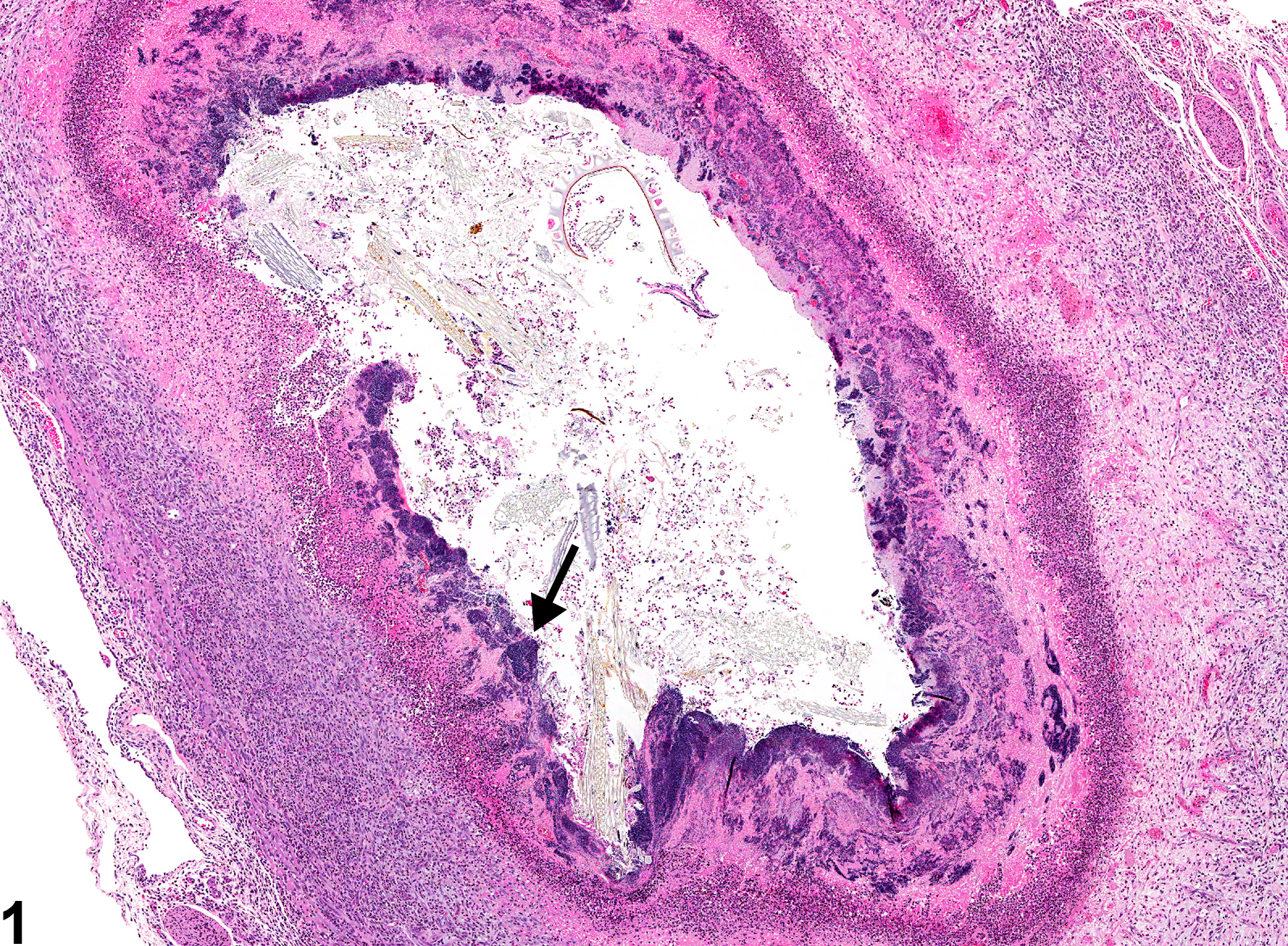
Esophagus - Inflammation, Suppurative in a female F344/N rat from a subchronic study. Note the bacterial colonies (arrow).
Figure 1 of 6
Image of inflammation in the esophagus from a female F344/N rat in a subchronic study
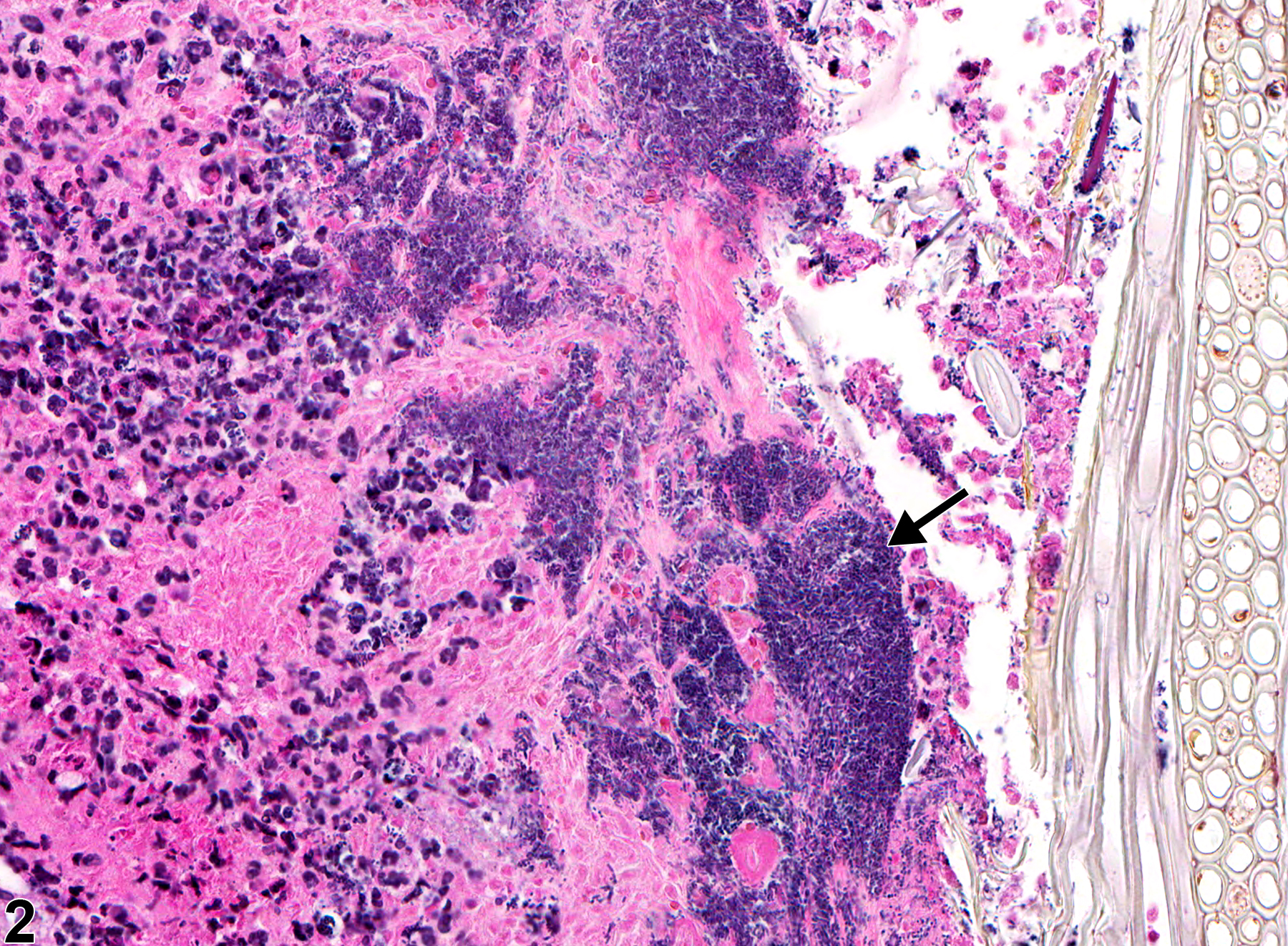
Esophagus - Inflammation, Suppurative in a female F344/N rat from a subchronic study (higher magnification of Figure 1). Note the bacterial colonies (arrow).
Figure 2 of 6
Image of inflammation in the esophagus from a male F344/N rat in a chronic study
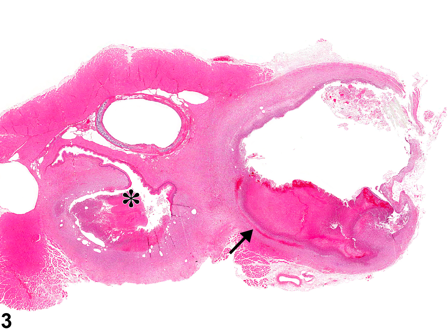
Esophagus - Inflammation, Suppurative in a male F344/N rat from a chronic study. There is inflammation in the esophagus (asterisk) and in the periesophageal tissue (arrow).
Figure 3 of 6
Image of inflammation in the esophagus from a male F344/N rat in a chronic study
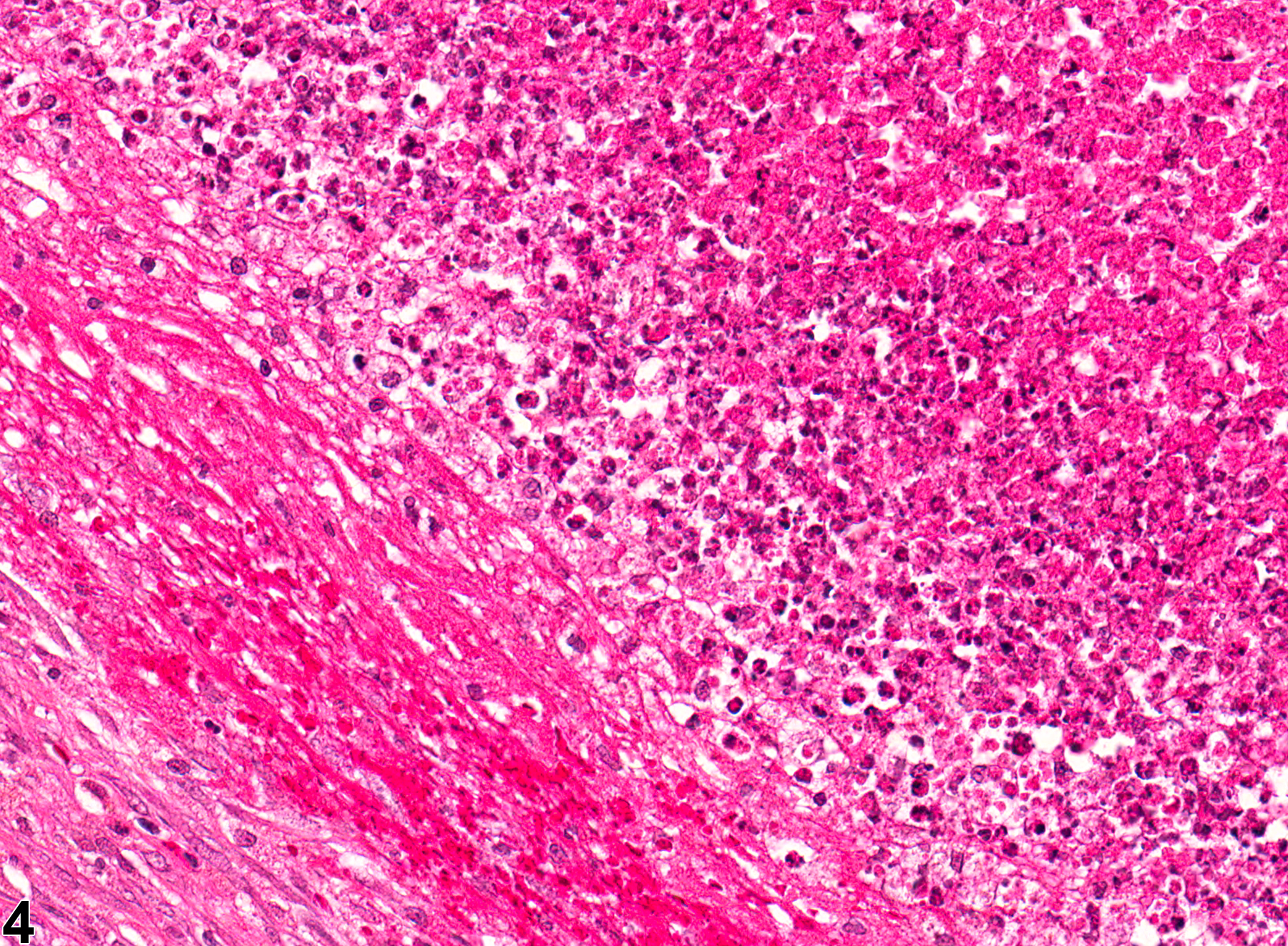
Esophagus - Inflammation, Suppurative in a male F344/N rat from a chronic study (higher magnification of Figure 3). Suppurative inflammation in the periesophageal tissue.
Figure 4 of 6
Image of inflammation in the esophagus from a female F344/N rat in a chronic study
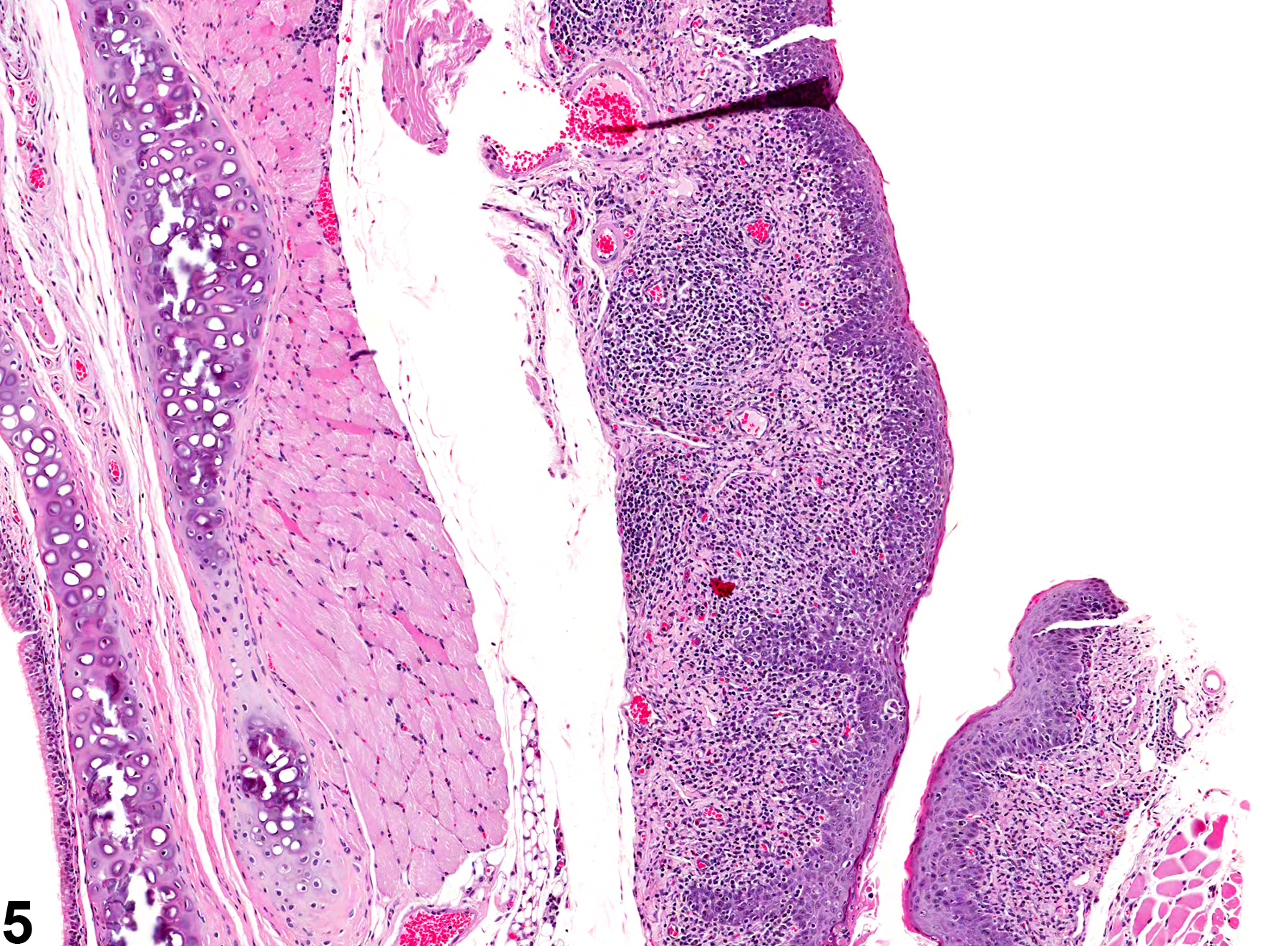
Esophagus - Inflammation, Chronic in a female F344/N rat from a chronic study. The inflammation is composed primarily of lymphocytes and plasma cells.
Figure 5 of 6
Image of inflammation in the esophagus from a female F344/N rat in a chronic study
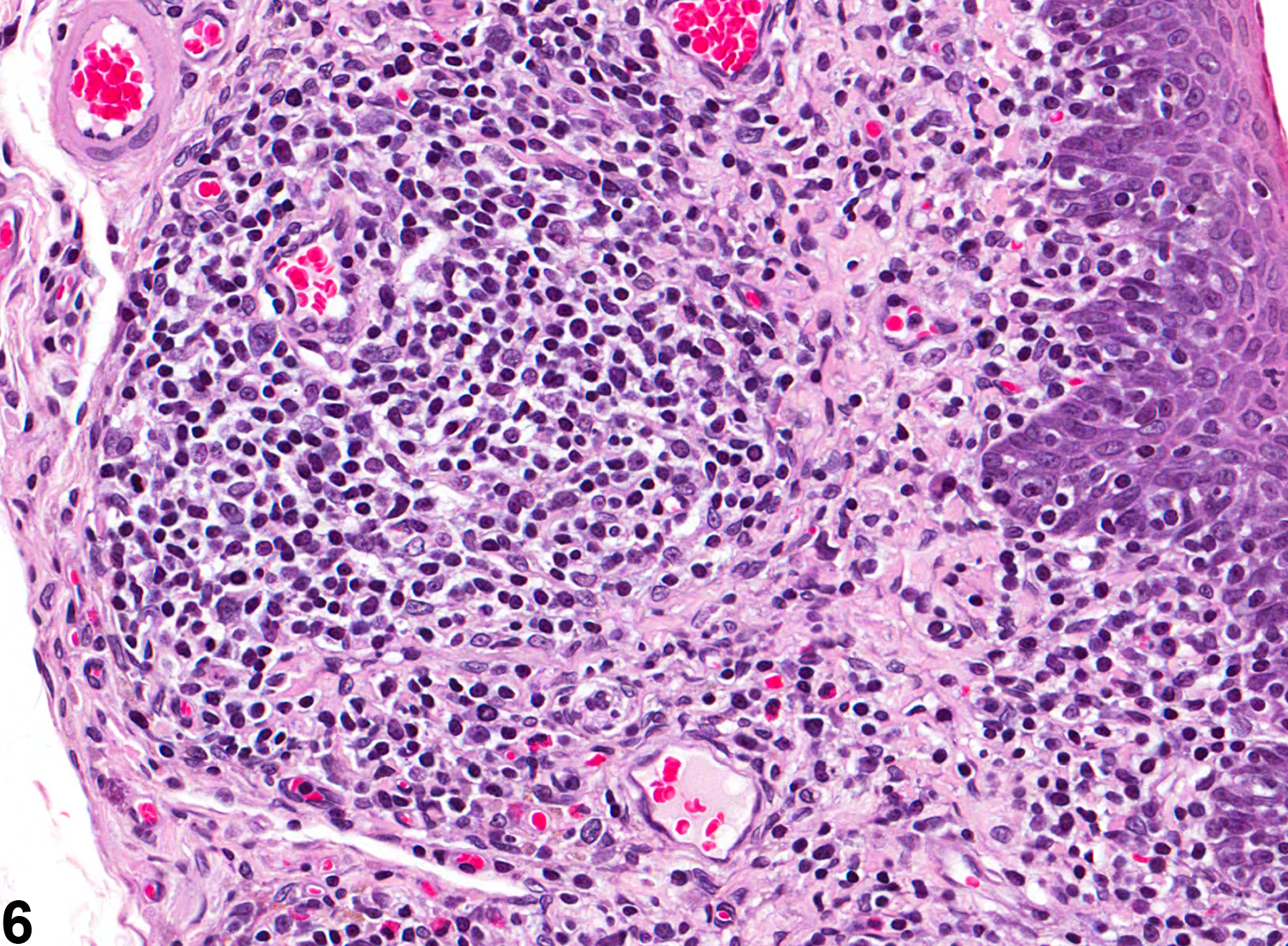
Esophagus - Inflammation, Chronic in a female F344/N rat from a chronic study (higher magnification of Figure 5). The inflammation is composed primarily of lymphocytes and plasma cells.
Figure 6 of 6
next arrow

comment:

Direct toxic effects of chemicals on the esophagus are rare in NTP studies. Esophageal inflammation in rodents is most commonly caused by gavage trauma and can be associated with ulceration, necrosis, and perforation. In NTP studies, there are five standard categories of inflammation: acute, suppurative, chronic, chronic active, and granulomatous. In acute inflammation, the predominant infiltrating cell is the neutrophil, though fewer macrophages and lymphocytes may also be present. There may also be evidence of edema or hyperemia. The neutrophil is also the predominant infiltrating cell type in suppurative inflammation, but the neutrophils are aggregated, and many of them are degenerate (suppurative exudate). The exudate may also contain cell debris, both from the resident cell populations and from infiltrating leukocytes; proteinaceous fluid containing fibrin; fewer macrophages; occasional lymphocytes or plasma cells; and, possibly, an infectious agent. Grossly, these lesions would be characterized by the presence of pus. In the tissue surrounding the exudate, there may be fibroblasts, fibrous connective tissue, and mixed inflammatory cells, depending on the chronicity of the lesion. Lymphocytes predominate in chronic inflammation. Lymphocytes also predominate in chronic active inflammation, but a significant number of neutrophils are also present. Both lesions may contain macrophages. Granulomatous inflammation is another form of chronic inflammation, but this diagnosis requires the presence of a significant number of aggregated, large, activated macrophages, epithelioid macrophages, or multinucleated giant cells. Suppurative inflammation with secondary bacterial infection due to gavage trauma is relatively common in the esophagus ( Figure 1image opens in a pop-up window , Figure 2image opens in a pop-up window , Figure 3image opens in a pop-up window , and Figure 4image opens in a pop-up window ).

recommendation:

Whenever present, inflammation should be diagnosed and graded. The diagnosis should include a modifier indicating the duration or type of inflammation (i.e., acute, suppurative, chronic, chronic active, or granulomatous). The severity grade depends on the extent of the lesion in the esophagus and the density of the cellular infiltrate. Lesions consistent with an abscess are diagnosed as suppurative inflammation. Associated necrosis or inflammation is not generally recorded unless the inflammation or necrosis is a significant component of the lesion.

references:

Ackermann MR. 2007. Acute inflammation. In: Pathologic Basis of Veterinary Disease, 4th ed (McGavin MD, Zachary JF, eds). Mosby, St Louis, MO, 101-152.

Ackermann MR. 2007. Chronic inflammation and wound healing. In: Pathologic Basis of Veterinary Disease, 4th ed (McGavin MD, Zachary JF, eds). Mosby, St Louis, MO, 153-191.

Brown HR, Hardisty JF. 1990. Oral cavity, esophagus and stomach. In: Pathology of the Fischer Rat (Boorman GA, Montgomery CA, MacKenzie WF, eds). Academic Press, San Diego, CA, 9-30.
Abstract: https://www.ncbi.nlm.nih.gov/nlmcatalog/9002563