

Endocrine System
Adrenal Gland - Fibrosis
Narrative
National Toxicology Program. 1997. NTP TR-459. Toxicology and Carcinogenesis Studies of t-Butylhydroquinone (CAS No. 1948-33-0) in F344/N Rats and B6C3F1 Mice (Feed Studies). NTP, Research Triangle Park, NC.
Abstract: https://ntp.niehs.nih.gov/go/6062National Toxicology Program. 1999. NTP TR-481. Toxicology and Carcinogenesis Studies of Oleic Acid Diethanolamine Condensate (CAS No. 93-83-4) in F344/N Rats and B6C3F1 Mice (Dermal Studies). NTP, Research Triangle Park, NC.
Abstract: https://ntp.niehs.nih.gov/go/9764National Toxicology Program. 2010. NTP TR-557. Toxicology and Carcinogenesis Studies of β-Myrcene (CAS No. 123-35-3) in F344/N Rats and B6C3F1 Mice (Gavage Studies). NTP, Research Triangle Park, NC.
Abstract: https://ntp.niehs.nih.gov/go/33584National Toxicology Program. 2010. NTP TR-559. Toxicology and Carcinogenesis Studies of 2,3',4,4',5-Pentachlorobiphenyl (PCB 118) (CAS No. 31508-00-6) in Female Harlan Sprague-Dawley Rats (Gavage Studies). NTP, Research Triangle Park, NC.
Abstract: https://ntp.niehs.nih.gov/go/33539National Toxicology Program. 2011. NTP TR-563. Toxicology and Carcinogenesis Studies of Pulegone (CAS No. 89-82-7) in F344/N Rats and B6C3F1 Mice (Gavage Studies). NTP, Research Triangle Park, NC.
Abstract: https://ntp.niehs.nih.gov/go/34851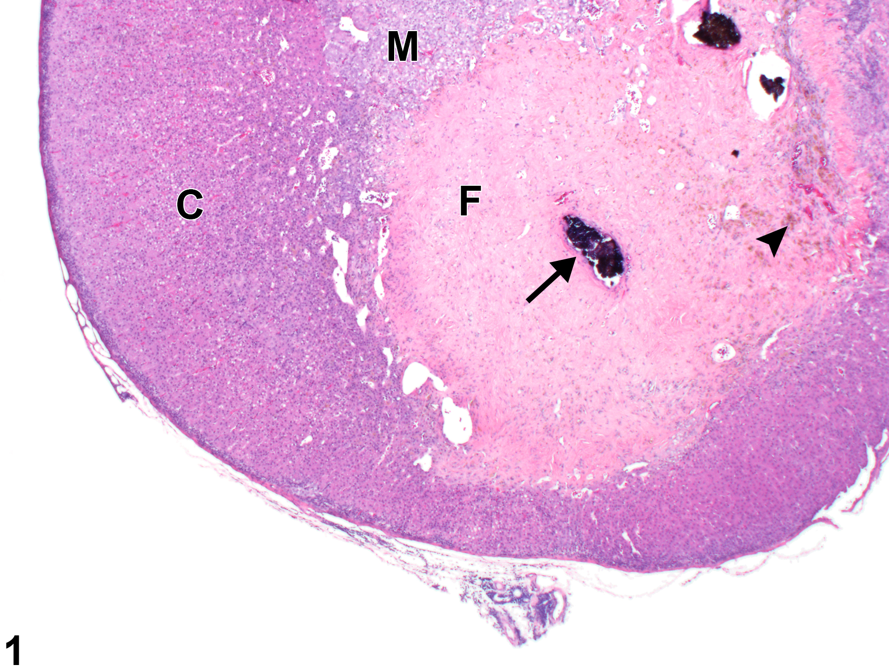
Adrenal gland - Fibrosis in a female F344/N rat from a chronic study. A large focal area of mature fibrous connective tissue (F) replaces parts of the normal cortex (C) and medulla (M); there is also focal mineralization (arrow) and a cluster of pigment-laden macrophages (arrowhead).
All Images

Adrenal gland - Fibrosis in a female F344/N rat from a chronic study. A large focal area of mature fibrous connective tissue (F) replaces parts of the normal cortex (C) and medulla (M); there is also focal mineralization (arrow) and a cluster of pigment-laden macrophages (arrowhead).

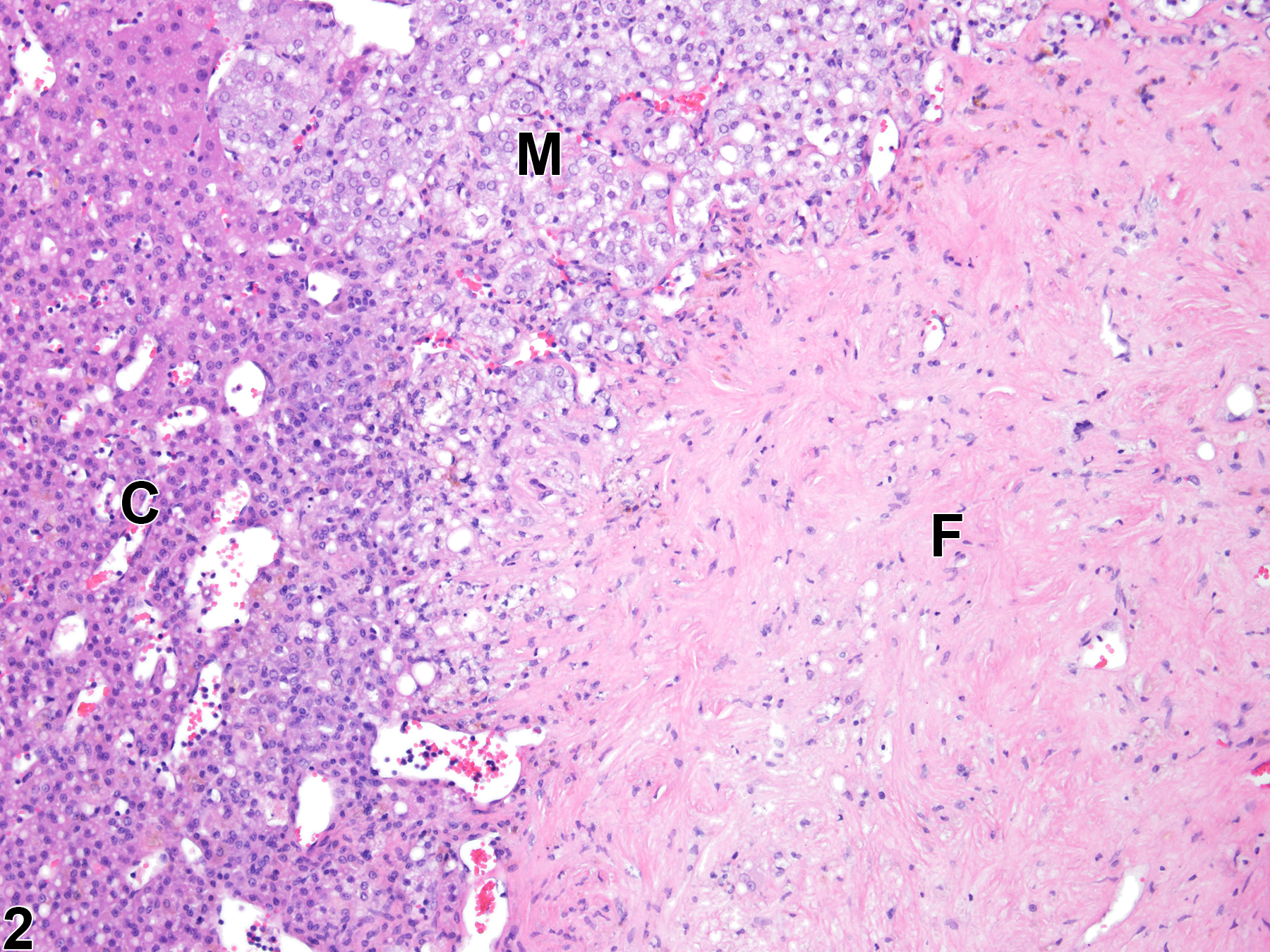
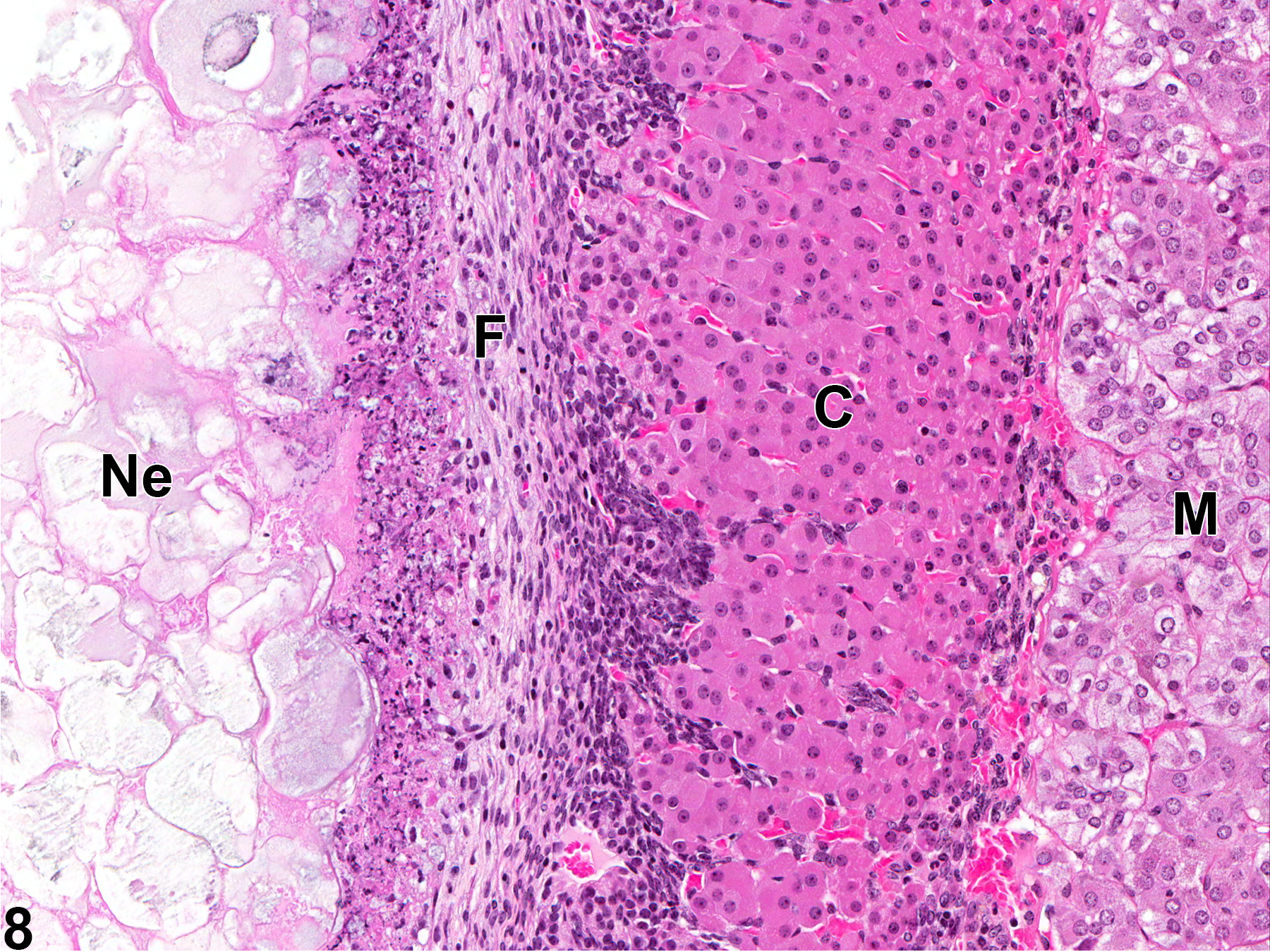
Adrenal gland - Fibrosis in a female F344/N rat from a chronic study (higher magnification of Figure 1). There are scattered fibroblasts and blood vessels within the focus of fibrosis (F); C = cortex, M = medulla.

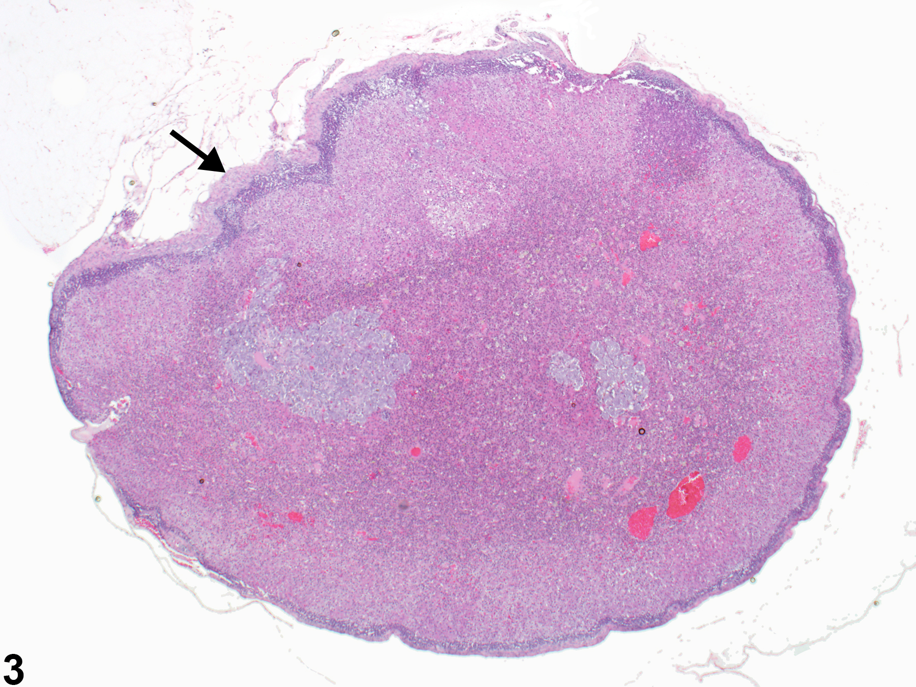
Adrenal gland, Capsule - Fibrosis in a female F344/N rat from a chronic study. Focal fibrosis (arrow) results in thickening, distortion, and undulation of the adrenal capsule.

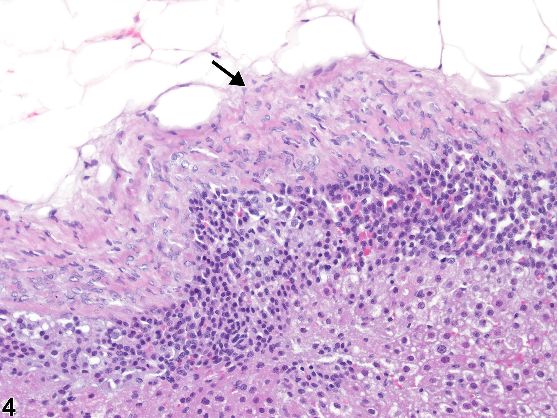
Adrenal gland, Capsule - Fibrosis in a female F344/N rat from a chronic study (higher magnification of Figure 3). Fibrosis of the adrenal capsule (arrow) is characterized by mature collagen sheets interspersed with fibrocytes.

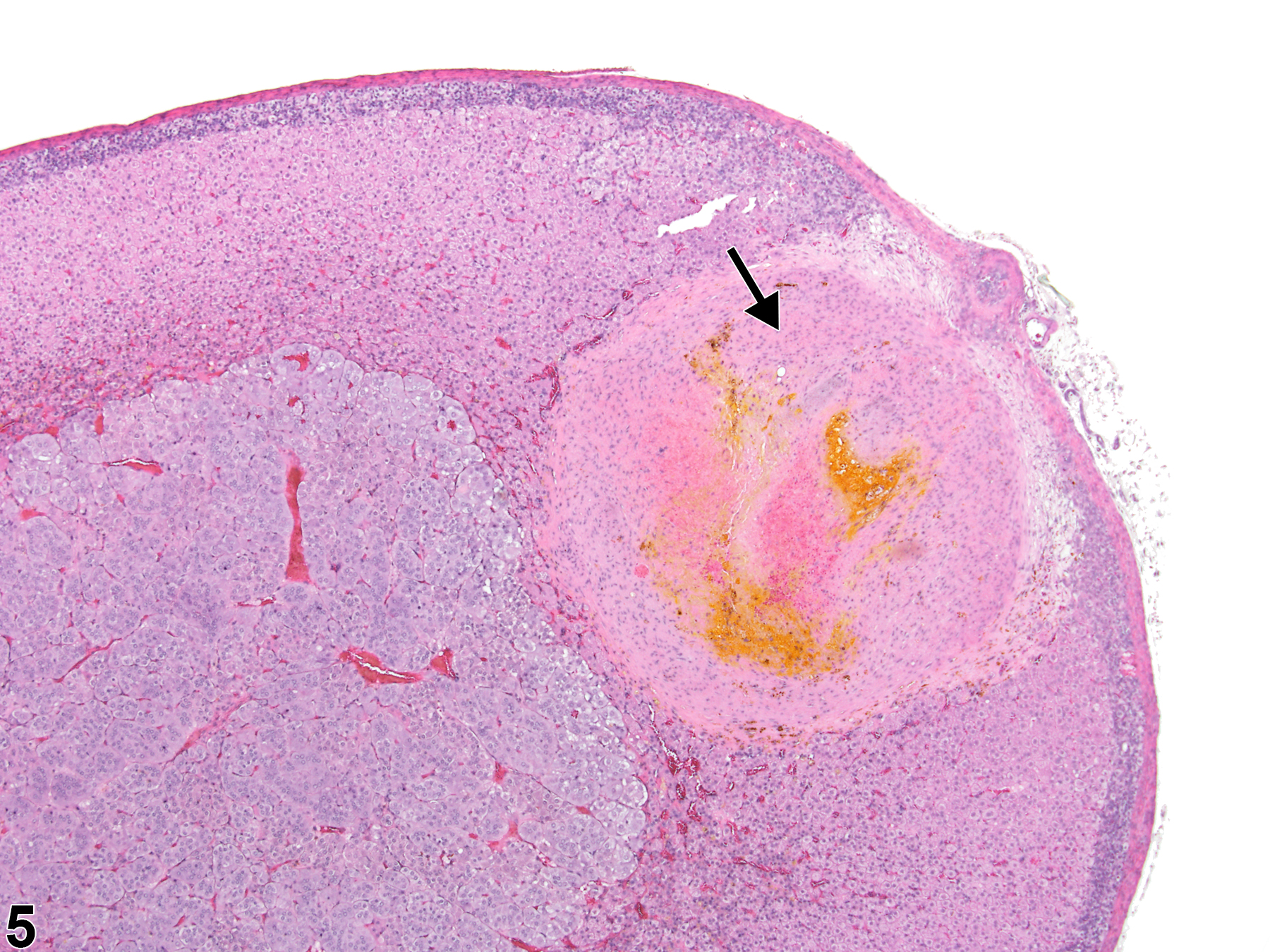
Adrenal gland, Cortex - Fibrosis in a female Sprague-Dawley rat from a chronic study. Focal fibrosis in the cortex is associated with an organized thrombus (arrow).

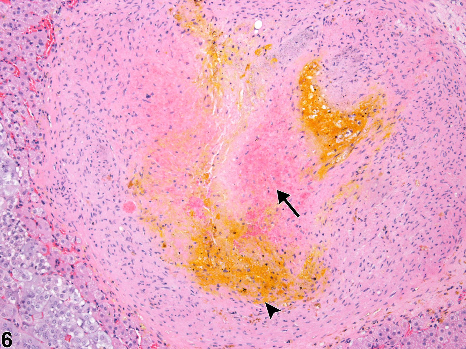
Adrenal gland, Cortex - Fibrosis in a female Sprague-Dawley rat from a chronic study (higher magnification of Figure 5). There is residual hemorrhage (arrow) and pigment accumulation (arrowhead) within the focus of fibrosis.

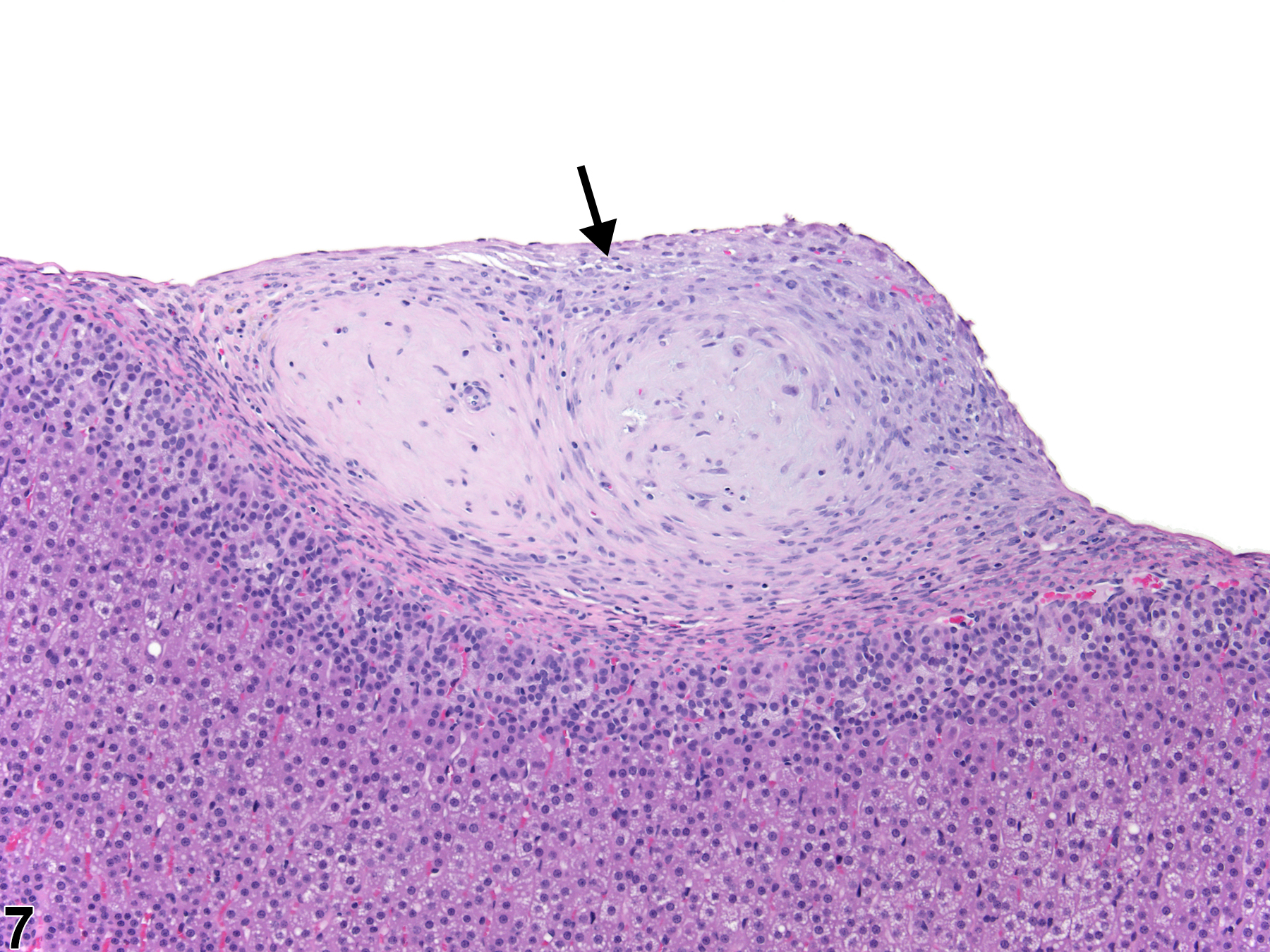
Adrenal gland, Capsule - Fibrosis in a male F344/N rat from a subchronic study. There is focal capsular fibrosis (arrow).

Adrenal gland, Capsule - Fibrosis in a female F344/N rat from a chronic study. The capsule is thickened by fibrosis (F), most likely a sequela to the necrosis (Ne) in the adjacent retroperitoneal fat. C = cortex, M = medulla.