

Endocrine System
Pituitary Gland
Narrative
Development of the pituitary gland involves an evagination from the dorsal surface of the oral cavity (craniopharyngeal or Rathke's pouch) toward the brain along with the formation of a downward vesicle from the floor of the brain toward the oral cavity. Eventually, the evagination forms the adenohypophysis, and the vesicle forms the neurohypophysis. The adenohypophysis consists of the pars distalis, pars tuberalis, and the pars intermedia. The pars distalis and pars tuberalis originate from the anterior wall of the Rathke's pouch; the pars tuberalis comes to form a sleeve-like covering of the infundibular stalk. The pars intermedia arises from a portion of the wall of the Rathke's pouch, which initially is in close proximity to the brain vesicle. Rathke's cleft is a remnant of Rathke's pouch that persists in the adult rat as a cleft or potential space between the pars intermedia and pars distalis. Rathke's cleft is typically obliterated in most species. In humans, Rathke's cleft is reduced to a small series of fluid-filled cysts with no known functional significance. Given the relatively small size of the rodent pituitary gland, routine handling, processing, and microtomy may not provide adequate histologic sampling of all components of the pituitary, especially when multiple tissues are embedded with the pituitary. In particular, the pars intermedia and nervosa may be partially or completely missed. The consistency or lack thereof in histologic sampling may affect evaluation of toxicity-related lesions and may necessitate additional sectioning of the pituitary tissue. If pituitary is a suspected target tissue, it may be prudent to embed it separately and consider taking multiple sections.
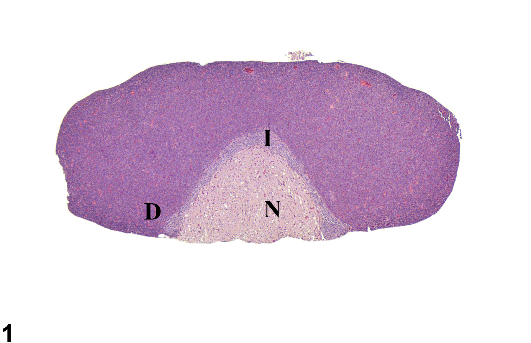
Figure 1. Pituitary Gland - Normal in a female F344/N rat from a chronic study. The normal pituitary consists of the pars distalis (D), the pars nervosa (N), and the pars intermedia (I).
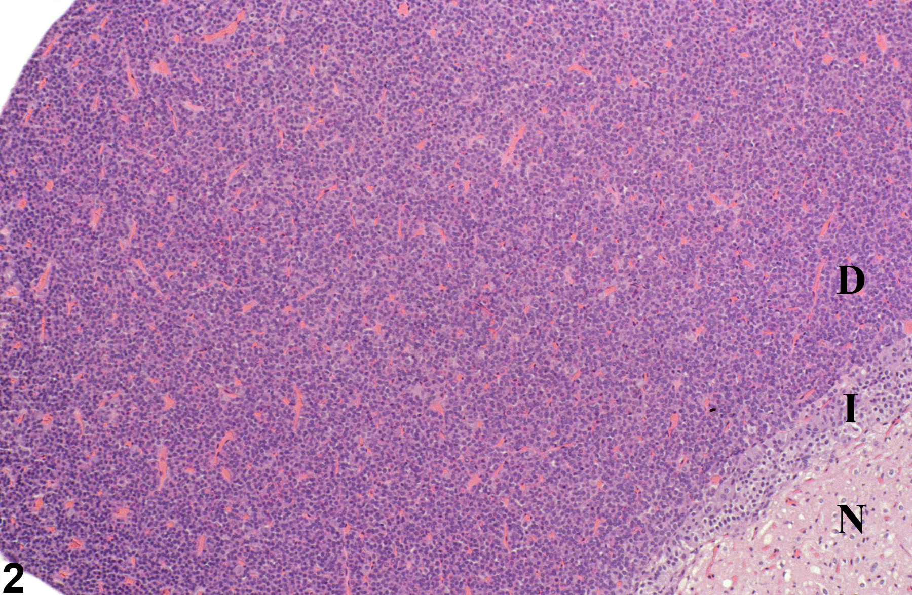
Figure 2. Pituitary Gland - Normal in a female F344/N rat from a chronic study. This higher magnification image of the normal pituitary shows the parts of the pituitary in higher detail.
MacKenzie WF, Boorman GA. 1991. Pituitary gland. In: Pathology of the Fischer Rat: Reference and Atlas (Boorman G, Eustis S, Elwell M, Montgomery CA, MacKenzie W, eds). Academic Press, San Diego, 485-500.
Abstract: http://www.ncbi.nlm.nih.gov/nlmcatalog/9002563Mahler J, Elwell M. 1999. The pituitary gland. In: Pathology of the Mouse: Reference and Atlas (Maronpot RR, Boorman GA, Gaul BW, eds). Cache River Press, Vienna, IL, 491-508.