

Endocrine System
Thyroid Gland, Follicle - Dilation
Narrative
Hardisty JF, Boorman GA. 1990. Thyroid gland. In: Pathology of the Fischer Rat: Reference and Atlas (Boorman GA, Eustis SL, Elwell MR, Montgomery CA, MacKenzie WF, eds). Academic Press, San Diego, 519–536.
Abstract: http://www.ncbi.nlm.nih.gov/nlmcatalog/9002563Hardisty JF, Boorman GA. 1999. Thyroid and parathyroid glands. In: Pathology of the Mouse: Reference and Atlas (Maronpot RR, Boorman GA, Gaul BW, eds). Cache River Press, Vienna, IL, 537–554.
Thyroid gland, Follicle - Dilation in a male B6C3F1 mouse from a chronic study. There is a single dilated follicle (arrow).
All Images

Thyroid gland, Follicle - Dilation in a male B6C3F1 mouse from a chronic study. There is a single dilated follicle (arrow).

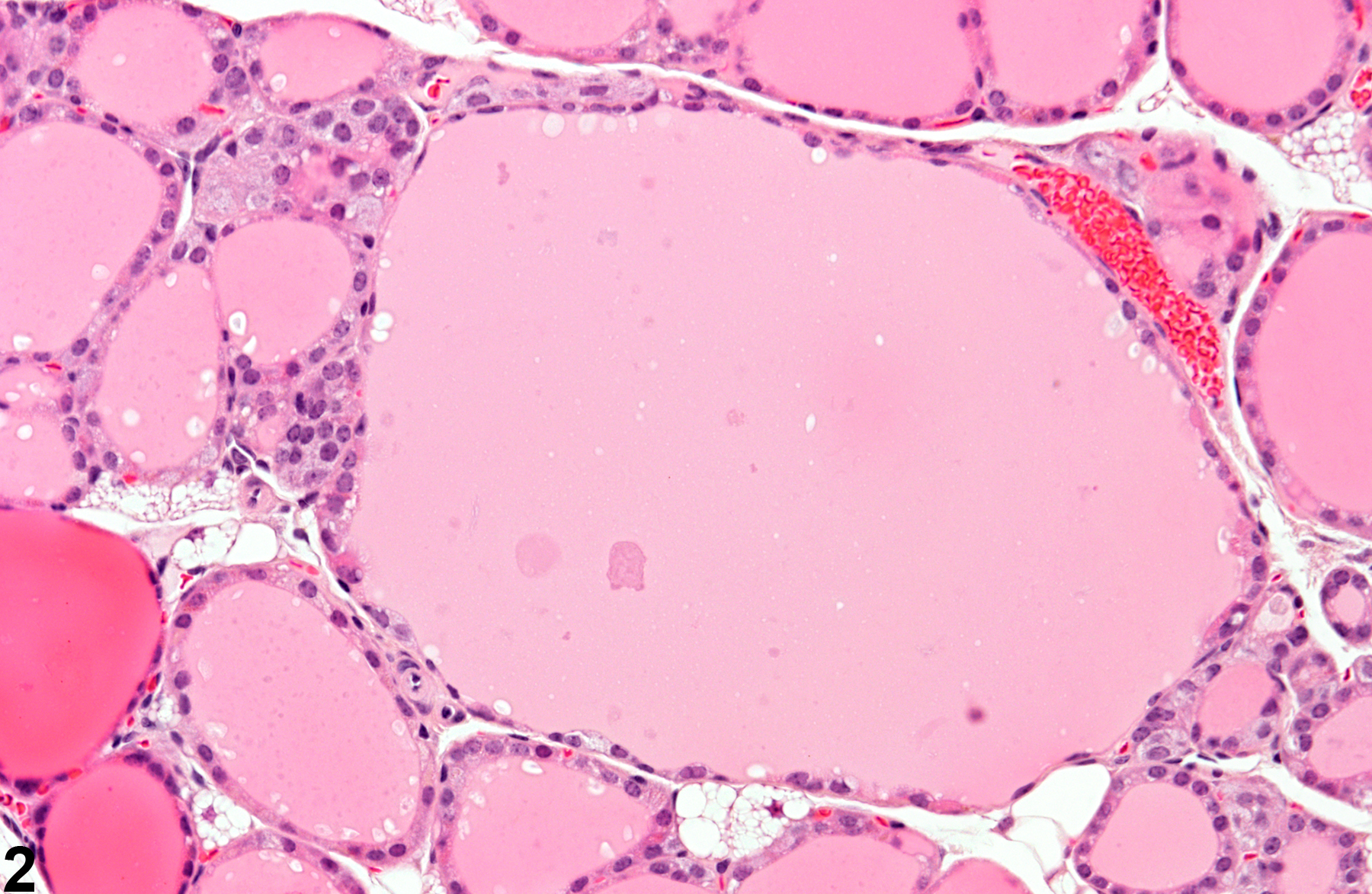
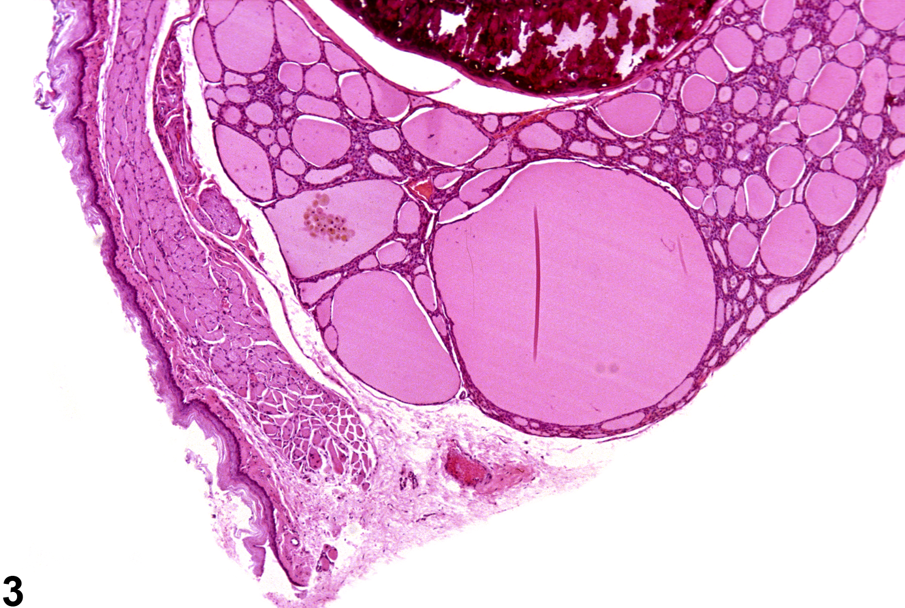
Thyroid gland, Follicle - Dilation in a male B6C3F1 mouse from a chronic study. This higher magnification of Figure 1 shows the flattened epithelium lining the dilated follicle.

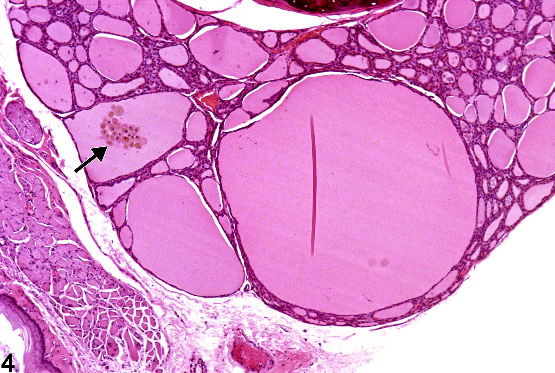
Thyroid gland, Follicle - Dilation in a male F344/N rat from a chronic study. There is dilation of several adjacent follicles.

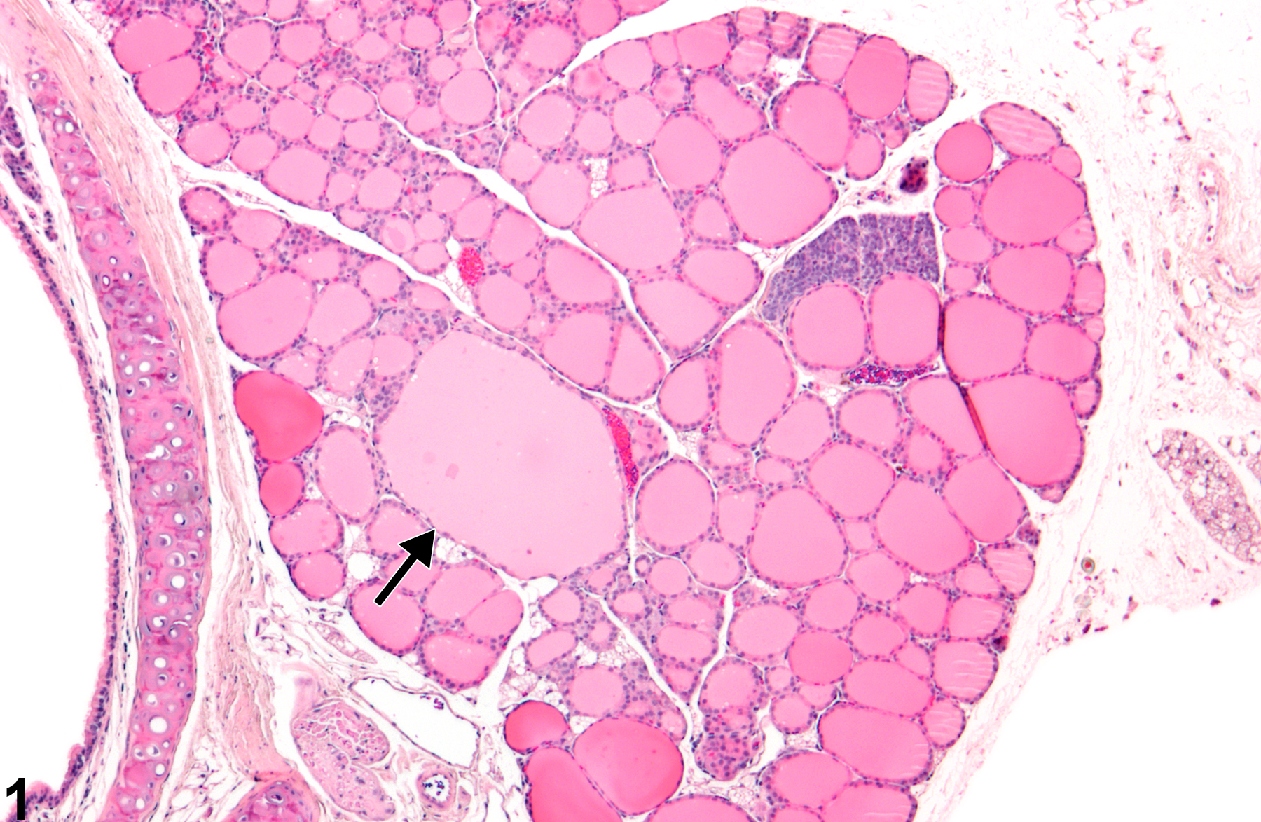
Thyroid gland, Follicle - Dilation in a male F344/N rat from a chronic study. Higher magnification of Figure 3 shows the flattened epithelium of the dilated follicles and pigmented macrophages in one of the follicles (arrow).