

Reproductive System, Female
Clitoral Gland - Infiltration Cellular
Narrative
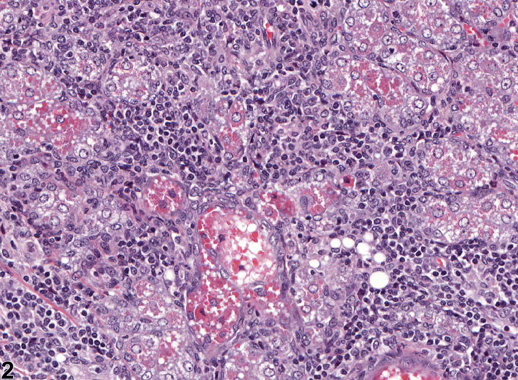
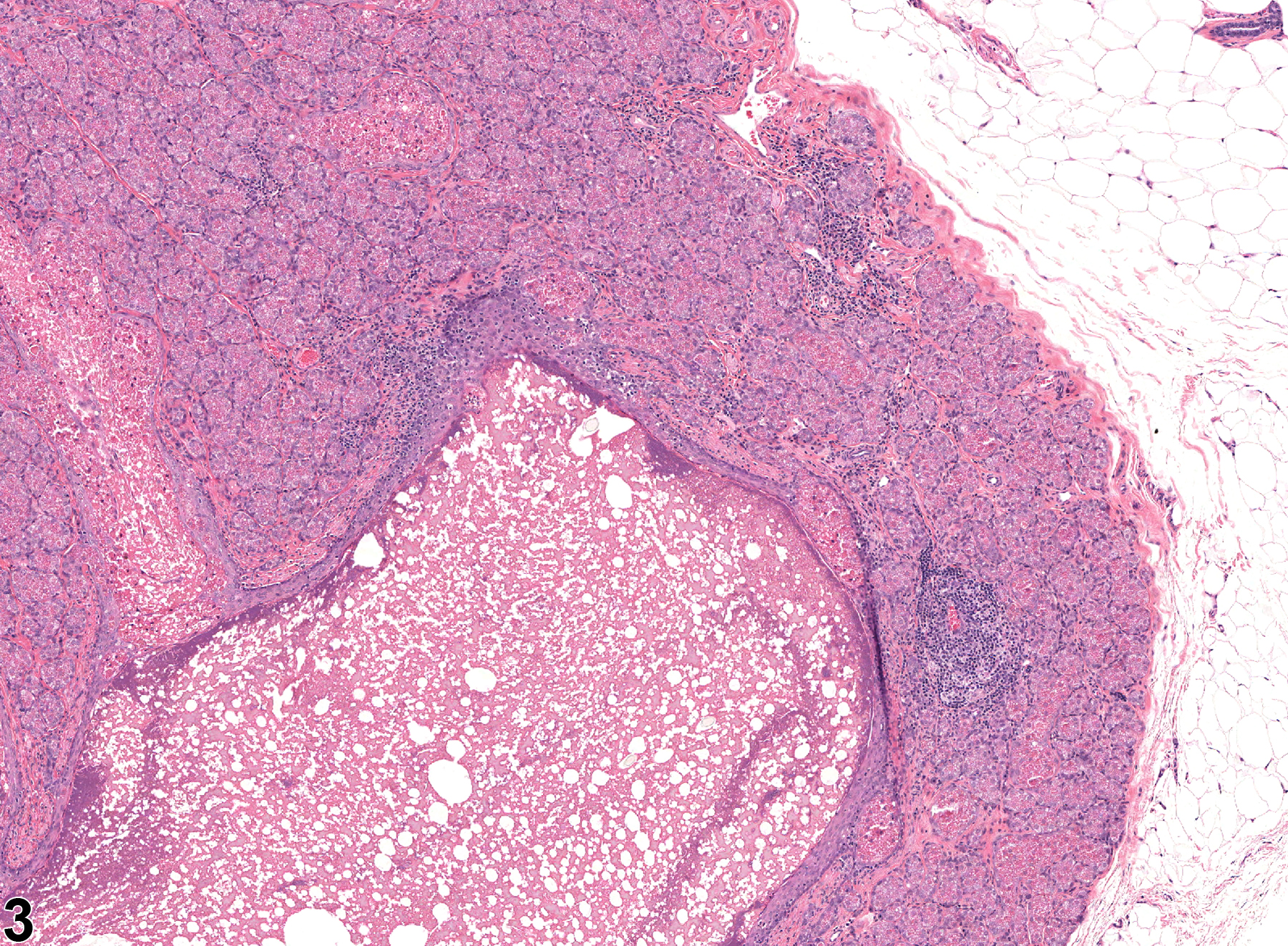
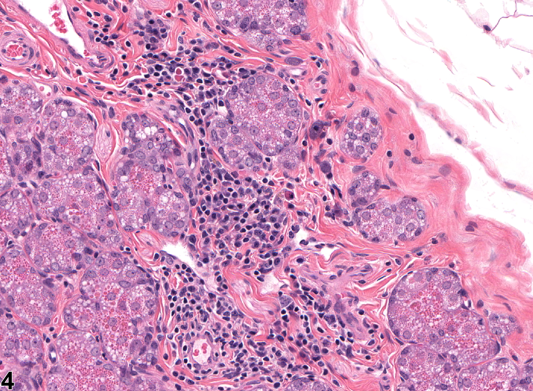
Clitoral gland - Infiltration, cellular may be a spontaneous age-related change in rats and mice but can also be a test-article-related effect. It is characterized by the presence of infiltrating cells (e.g., mononuclear cells, neutrophils, mast cells) within the tissue but with no other evidence of inflammation, such as tissue damage, necrosis, etiologic agents, hemorrhage, congestion, or edema (Figure 1, Figure 2, Figure 3 and Figure 4). Infiltration cellular must be differentiated from inflammation, in which there is other evidence of inflammation as listed above.
Clitoral gland - Infiltration cellular should be diagnosed and assigned a severity grade. The type of infiltrating cell should be specified as a modifier in the diagnosis.
Copeland-Haines D, Eustis SL. 1990. Specialized sebaceous glands. In: Pathology of the Fischer Rat: Reference and Atlas (Boorman GA, Eustis SL, Elwell MR, Montgomery CA, MacKenzie WF, eds). Academic Press, San Diego, CA, 279-293.
National Toxicology Program. 2011. NTP TR-570. Toxicology and Carcinogenesis Studies of α,β-Thujone (CAS No. 76231-76-0) in F344/N Rats and B6C3F1 Mice (Gavage Studies). NTP, Research Triangle Park, NC.
Abstract: https://ntp.niehs.nih.gov/go/36137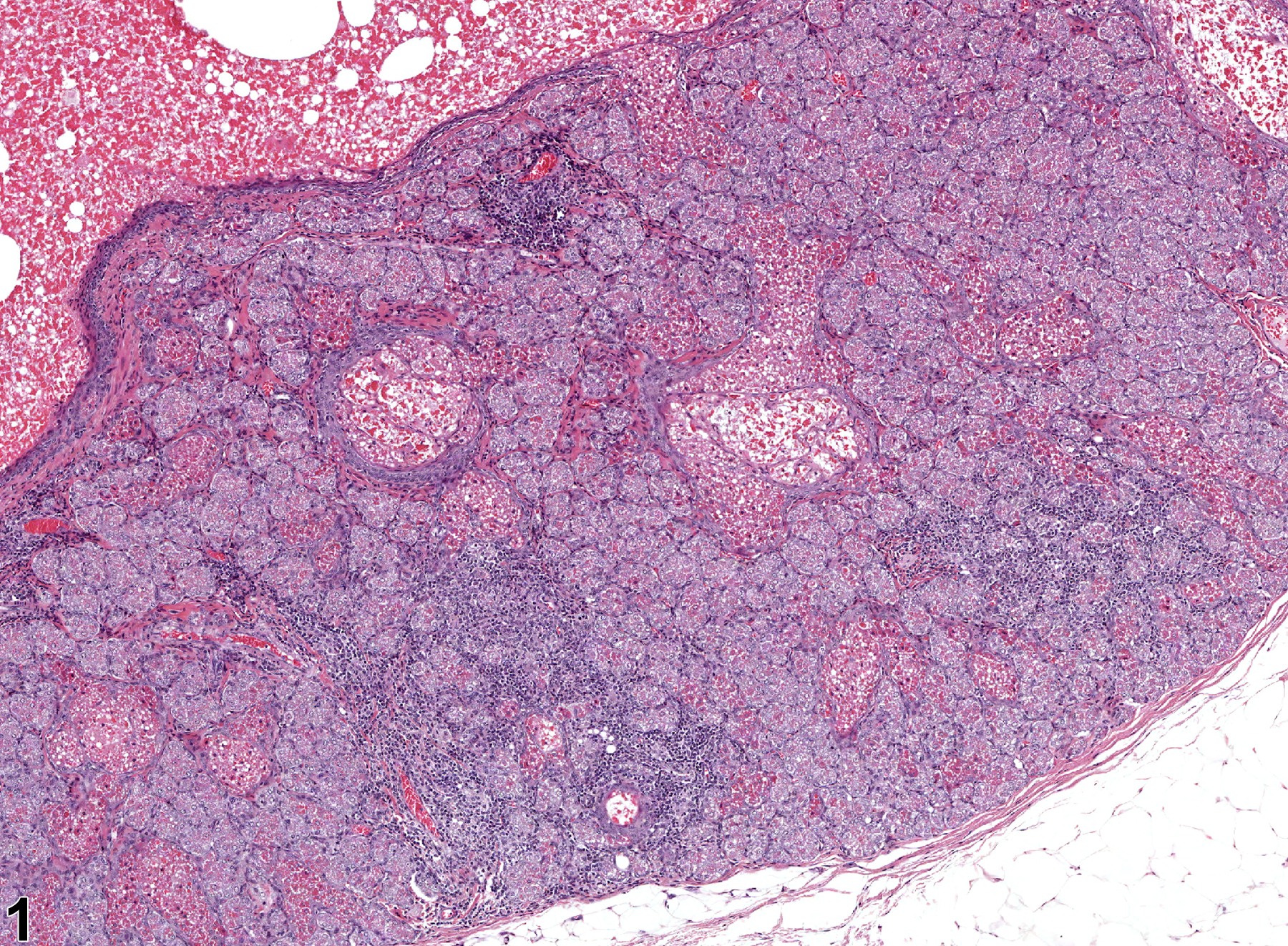
Clitoral gland - Infiltration cellular, Lymphocyte and Plasma cells in a female F344/N rat from a chronic study. An infiltration of lymphocytes and plasma cells is present in the stroma.
All Images

Clitoral gland - Infiltration cellular, Lymphocyte and Plasma cells in a female F344/N rat from a chronic study. An infiltration of lymphocytes and plasma cells is present in the stroma.

Clitoral gland - Infiltration cellular, Lymphocytes and Plasma cells in a female F344/N rat from a chronic study (higher magnification of Figure 1). A stromal cellular infiltrate of lymphocytes and plasma cells is present.

Clitoral gland - Infiltration cellular, Lymphocyte in a female F344/N from a subchronic study. Lymphocytic infiltration is present.

Clitoral gland - Infiltration cellular, Lymphocyte in a female F344/N from a subchronic study (higher magnification of Figure 3). Lymphocytic infiltration is present in the stroma.