

Reproductive System, Female
Ovary - Fatty Change
Narrative
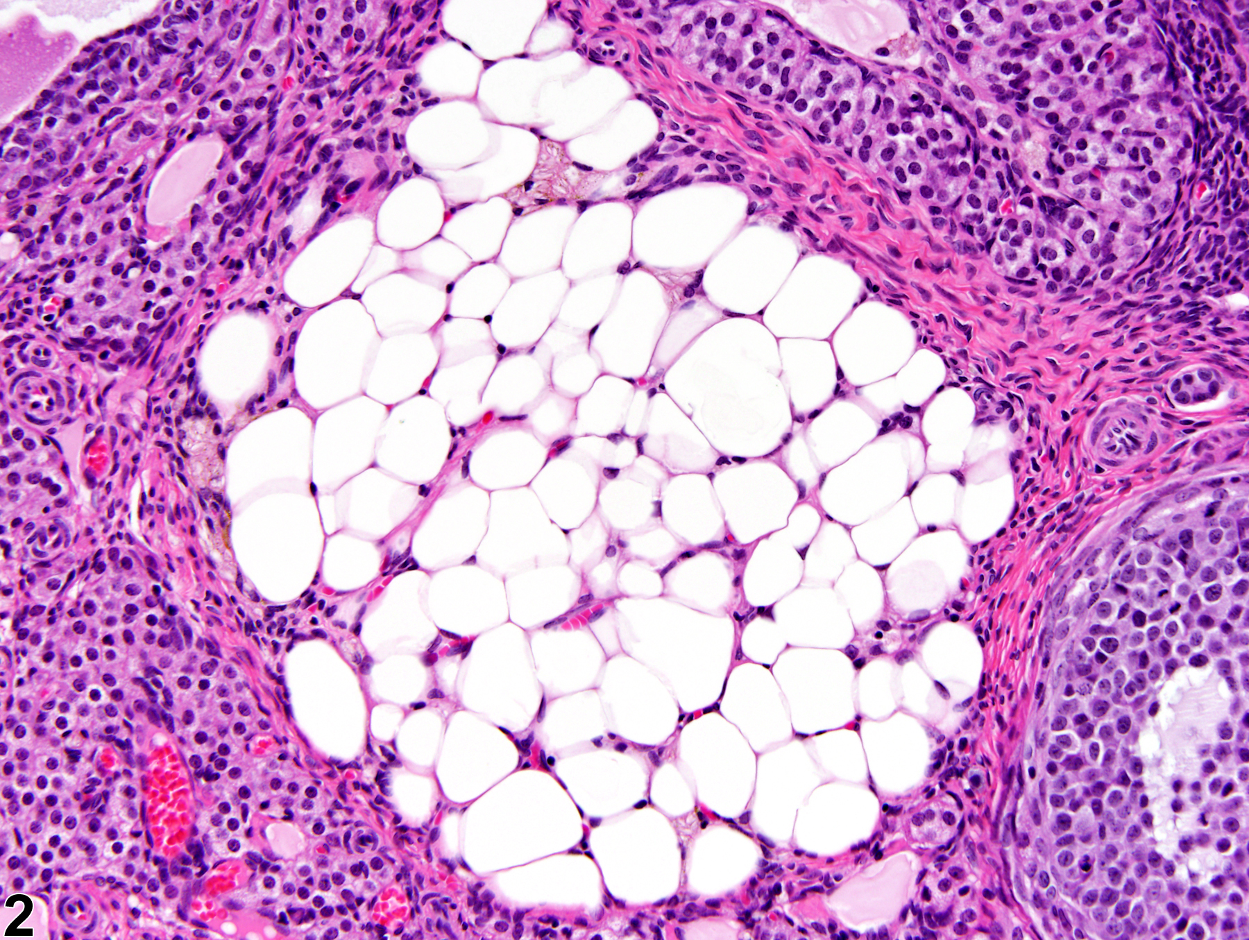
Ovary - Fatty change is characterized by a well-circumscribed, partially encapsulated, slightly compressing cluster of cells with the appearance typical of adipocytes (Figure 1 and Figure 2). Large, variably sized, clear vacuoles are present in the cytoplasm of polygonal cells, displacing the flattened nucleus to the periphery of the cell. Interstitial, thecal, and granulosa cells contain numerous lipid droplets. Fatty change may be induced by drugs inhibiting steroid hormone synthesis, resulting in lipid accumulation in the ovarian stroma. Rats treated with an imidazole antifungal drug had increased lipid in the interstitial cells of the ovary, which was also associated with deposition of lipofuscin.
Ovary - Fatty change should be diagnosed and graded whenever present.
Alison RH, Morgan KT, Montgomery CA. 1990. Ovary. In: Pathology of the Fischer Rat: Reference and Atlas (Boorman GA, Eustis SL, Elwell MR, Montgomery CA, MacKenzie WF, eds). Academic Press, San Diego, CA, 429-442.
Greaves P. 2012. Female genital tract. In: Histopathology of Preclinical Toxicity Studies: Interpretation and Relevance in Drug Safety Evaluation, 4th ed. Elsevier, Amsterdam, 667-724.
Peluso JJ, Gordon LR. 1992. Nonneoplastic and neoplastic changes in the ovary. In: Pathobiology of the Aging Rat (Mohr U, Dungworth DL, Capen CC, eds). ILSI Press, Washington, DC, 351-364.
Ovary - Fatty change in a female F344/N rat from a subchronic study. There is focal accumulation of adipocytes in the ovarian parenchyma.
All Images

Ovary - Fatty change in a female F344/N rat from a subchronic study. There is focal accumulation of adipocytes in the ovarian parenchyma.

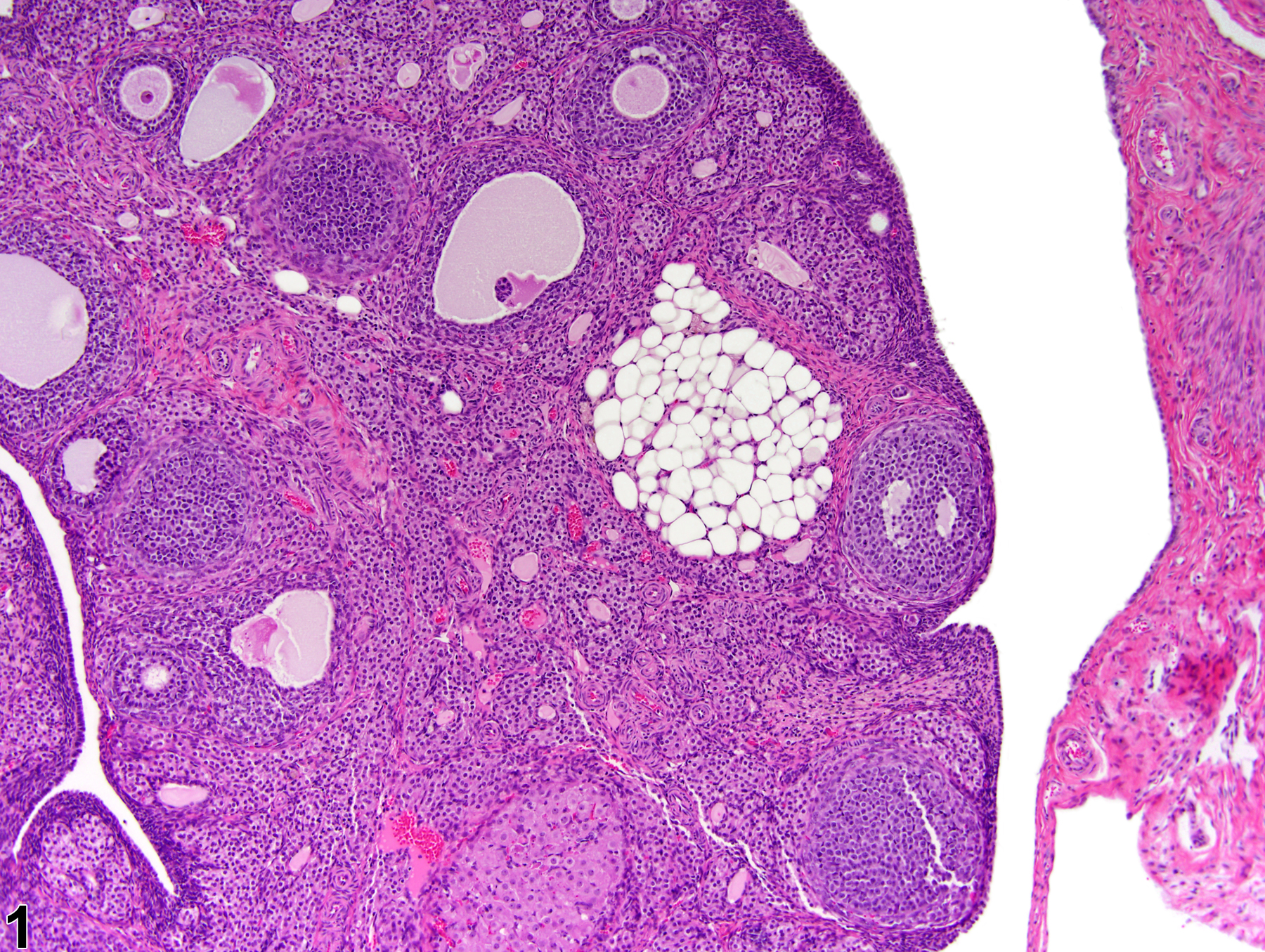
Ovary - Fatty change in a female F344/N rat from a subchronic study (higher magnification of Figure 1). There is localized adipose tissue accumulation in ovarian parenchyma.