

Hepatobiliary System
Gallbladder - Hyaline Droplet Accumulation
Narrative
Harada T, Enomoto A, Boorman GA, Maronpot RR. 1999. Liver and gallbladder. In: Pathology of the Mouse: Reference and Atlas (Maronpot RR, Boorman GA, Gaul BW, eds). Cache River Press, Vienna, IL, 119-183.
Hsu L, Diwan B, Ward JM, Noguchi CT. 2006. Pathology of ‘‘Berkeley’’ sickle-cell mice includes gallstones, priapism. Blood 107:3414-3415.
Abstract: http://www.ncbi.nlm.nih.gov/pubmed/16597602Thoolen B, Maronpot RR, Harada T, Nyska A, Rousseaux C, Nolte T, Malarkey D, Kaufmann W, Kutter K, Deschl U, Nakae D, Gregson R, Winlove M, Brix A, Singl B, Belpoggi F, Ward JM. 2010. Hepatobiliary lesion nomenclature and diagnostic criteria for lesions in rats and mice (INHAND). Toxicol Pathol 38:5S-81S.
Full Text: http://tpx.sagepub.com/content/38/7_suppl/5S.fullWard JM, Yoon M, Anver MR, Haines DC, Kudo G, Gonzalez FJ, Kimura S. 2001. Hyalinosis and Ym1/Ym2 gene expression in the stomach and respiratory tract of 129S4/SvJae and wild-type and CYP1A2-null B6, 129 mice. Am J Pathol 158:323-332.
Abstract: http://www.ncbi.nlm.nih.gov/pubmed/11141507Yang YH, Campbell JS. 1964. Crystalline excrements in bronchitis and cholecystitis of mice. Am J Pathol 45:337-345.
Abstract: http://www.ncbi.nlm.nih.gov/pmc/articles/PMC1907179/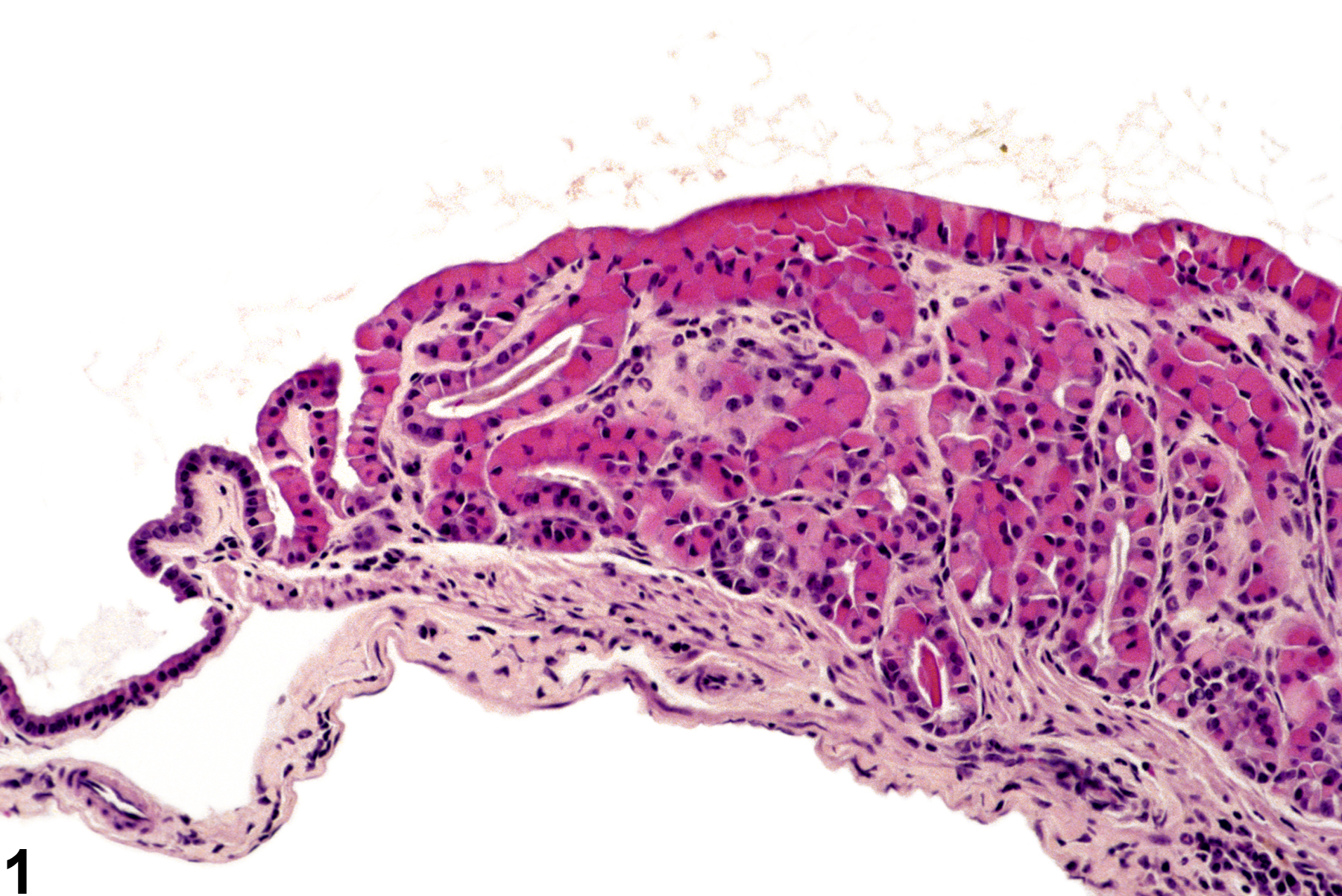
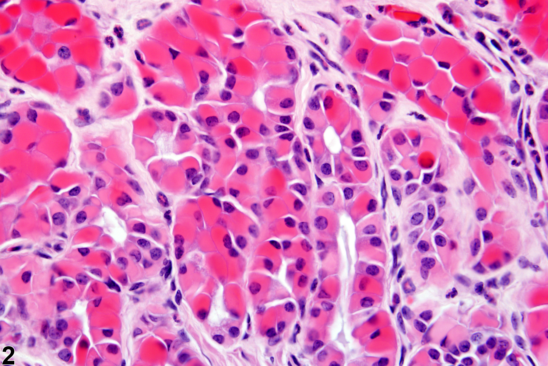
Hyaline droplet accumulation in the gallbladder in a male B6C3F1 mouse from a chronic study.
All Images

Hyaline droplet accumulation in the gallbladder in a male B6C3F1 mouse from a chronic study.

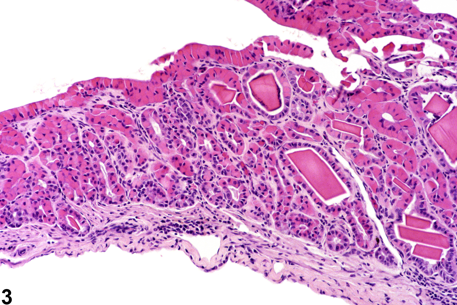
Hyaline droplet accumulation in the gallbladder in a male B6C3F1 mouse from a chronic study (higher magnification of Figure 1).

Hyaline droplet accumulation with extracellular crystalline material in the gallbladder in a male B6C3F1 mouse from a chronic study.