

Hepatobiliary System
Liver - Hepatodiaphragmatic Nodule
Narrative
Eustis SL, Boorman GA, Harada T, Popp JA. 1990. Liver. In: Pathology of the Fischer Rat (Boorman GA, Eustis SL, Elwell MR, Montgomery CA, MacKenzie WF, eds). Academic Press, San Diego, 71-94.
National Toxicology Program. 1982. NTP TR-228. Carcinogenesis Bioassay of Vinylidene Chloride (CAS No. 75-35-4) in F344/N Rats and B6C3F1/N Mice (Gavage Study). NTP, Research Triangle Park, NC.
Full Text: https://ntp.niehs.nih.gov/ntp/htdocs/lt_rpts/tr228.pdfThoolen B, Maronpot RR, Harada T, Nyska A, Rousseaux C, Nolte T, Malarkey D, Kaufmann W, Kutter K, Deschl U, Nakae D, Gregson R, Winlove M, Brix A, Singl B, Belpoggi F, Ward JM. 2010. Hepatobiliary lesion nomenclature and diagnostic criteria for lesions in rats and mice (INHAND). Toxicol Pathol 38:5S-81S.
Full Text: http://tpx.sagepub.com/content/38/7_suppl/5S.full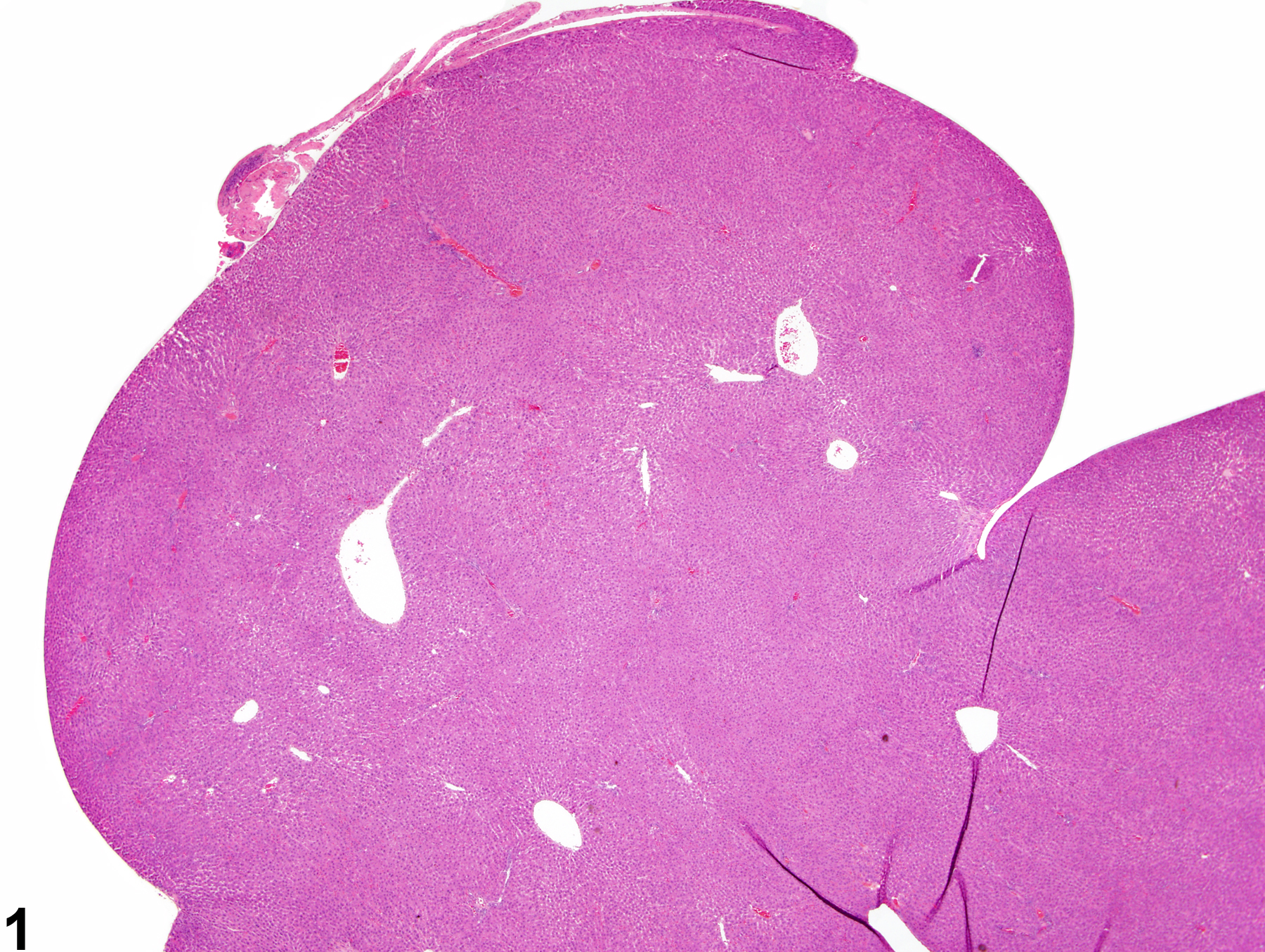
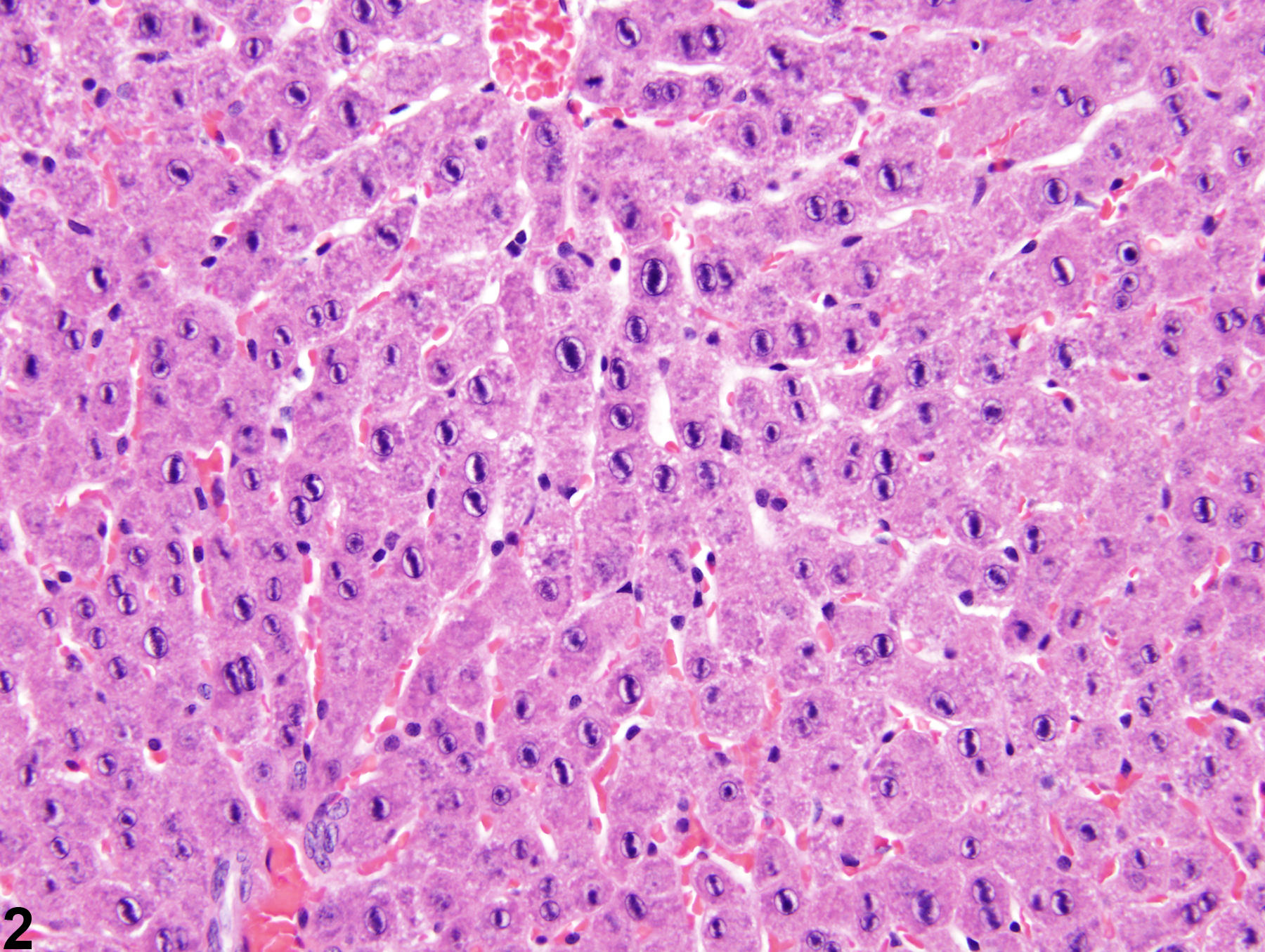
Hepatodiaphragmatic nodule in a female F344/N rat from a subchronic study.
All Images

Hepatodiaphragmatic nodule in a female F344/N rat from a subchronic study.

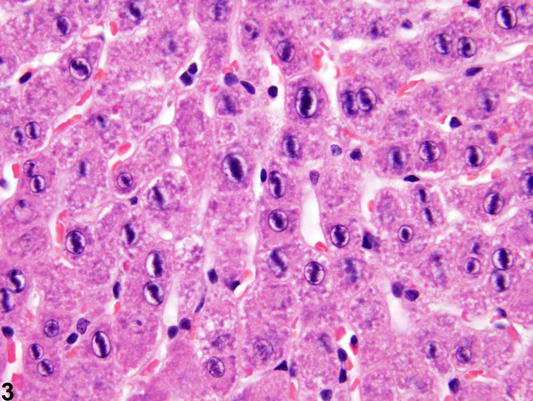
Hepatodiaphragmatic nodule in a female F344/N rat from a subchronic study. Note the linear chromatin arrangement in the nuclei.

Hepatodiaphragmatic nodule in a female F344/N rat from a subchronic study. Note the linear chromatin arrangement in the nuclei (higher magnification of Figure 2).