U.S. flag

An official website of the United States government

Dot gov

The .gov means it's official.
Federal government websites often end in .gov or .mil. Before sharing sensitive information, make sure you're on a federal government site.

Https

The site is secure.
The https:// ensures that you are connecting to the official website and that any information you provide is encrypted and transmitted securely.

Skip to Main Navigation
Skip to Page Content
Skip to Atlas Navigation

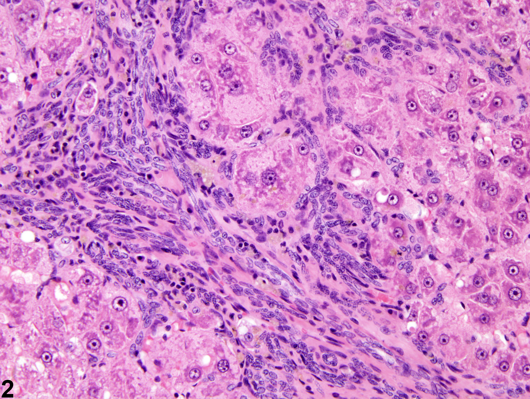
Liver, Hepatocyte – Hyperplasia, Nodular

Image of hyperplasia, nodular in the liver from a female Harlan Sprague-Dawley rat in a chronic study
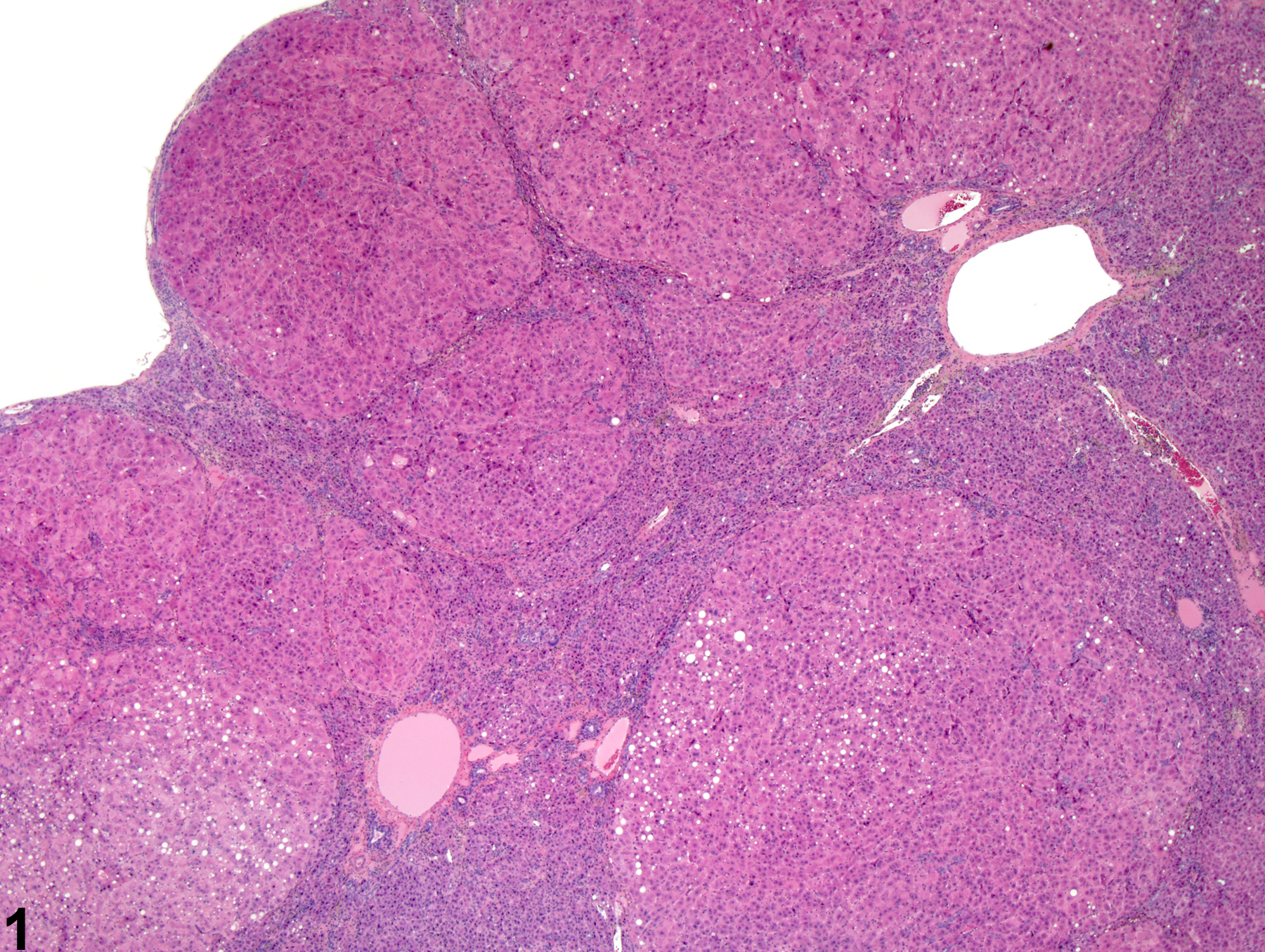
Nodular hyperplasia in a female Harlan Sprague-Dawley rat from a chronic study.
Figure 1 of 2
Image of hyperplasia, nodular in the liver from a female Harlan Sprague-Dawley rat in a chronic study
Nodular hyperplasia in a female Harlan Sprague-Dawley rat from a chronic study (higher magnification of Figure 1).
Figure 2 of 2
next arrow

comment:

Nodular hyperplasia can occur as a sequela to chronic hepatotoxicity or may arise in an otherwise normal liver. It may also be secondary to large granular cell lymphoma (mononuclear cell leukemia). It usually consists of multiple nodules of hepatocellular hyperplasia separated by bands of oval cells, collagen deposition, and collapsed hepatic lobules ( Figure 1image opens in a pop-up window and Figure 2image opens in a pop-up window ). Varying numbers of mononuclear leukocytes may also be present. Nodular hyperplasia results in gross distortion of the liver with retention of a markedly distorted hepatic lobular structure. Evidence of current or history of antecedent hepatotoxicity indicates a regenerative pathogenesis.

recommendation:

Whenever present, this chronic, proliferative, hepatic response should be diagnosed and given a severity grade, with severity dependent on the extent of liver involvement. Associated lesions, such as inflammation, fibrosis, or hepatocellular necrosis, should be diagnosed separately if warranted by the severity of these lesions. Their association can be made clear in the pathology narrative. Current or antecedent hepatotoxicity should be commented on in the pathology narrative.

references:

Eustis SL, Boorman GA, Harada T, Popp JA. 1990. Liver. In: Pathology of the Fischer Rat (Boorman GA, Eustis SL, Elwell MR, Montgomery CA, MacKenzie WF, eds). Academic Press, San Diego, 71-94.

Hailey JR, Walker NJ, Sells DM, Brix AE, Jokinen MP, Nyska A. 2005. Classification of proliferative hepatocellular lesions in Harlan Sprague-Dawley rats chronically exposed to dioxin-like compounds. Toxicol Pathol 33:165-174.
Abstract: https://www.ncbi.nlm.nih.gov/pubmed/15805068

Thoolen B, Maronpot RR, Harada T, Nyska A, Rousseaux C, Nolte T, Malarkey D, Kaufmann W, Kutter K, Deschl U, Nakae D, Gregson R, Winlove M, Brix A, Singl B, Belpoggi F, Ward JM. 2010. Hepatobiliary lesion nomenclature and diagnostic criteria for lesions in rats and mice (INHAND). Toxicol Pathol 38:5S-81S.
Full Text: http://tpx.sagepub.com/content/38/7_suppl/5S.full