

Immune System
Spleen - Atrophy
Narrative
Elmore SA. 2006. Enhanced histopathology of the spleen. Toxicol Pathol 34:648-655.
Full Text: https://www.ncbi.nlm.nih.gov/pmc/articles/PMC1828535/National Toxicology Program. 1992. 13-Week Short-Term Study of A3'-Azido-3'-Deoxythymidine (AZT) + Methadone Hydrochloride in B6C3F1 Mice (Gavage Studies) Completed (C92014). NTP, Research Triangle Park, NC.
National Toxicology Program. 2002. NTP TR-507. Toxicology and Carcinogenesis Studies of Vanadium Pentoxide (CAS No. 1314-62-1) in F344/N Rats and B6C3F1 Mice (Inhalation Studies). NTP, Research Triangle Park, NC.
Abstract: https://ntp.niehs.nih.gov/go/14892Stefanski SA, Elwell MR, Stromberg PC. 1990. Spleen, lymph nodes, and thymus. In: Pathology of the Fischer Rat: Reference and Atlas (Boorman GA, Eustis SL, Elwell MR, Montgomery CA, MacKenzie WF, eds). Academic Press, San Diego, 369-394.
Suttie AW. 2006. Histopathology of the spleen. Toxicol Pathol 34:466-503.
Full Text: http://tpx.sagepub.com/content/34/5/466.full.pdfWard JM, Mann PC, Morishima H, Frith CH. 1999. Thymus, spleen, and lymph nodes. In: Pathology of the Mouse (Maronpot RR, ed). Cache River Press, Vienna, IL, 333-360.
Ward JM, Rehg JE, Morse HC III. 2012. Differentiation of rodent immune and hematopoietic system reactive lesions from neoplasias. Toxicol Pathol 40:425-434.
Abstract: https://www.ncbi.nlm.nih.gov/pubmed/22215512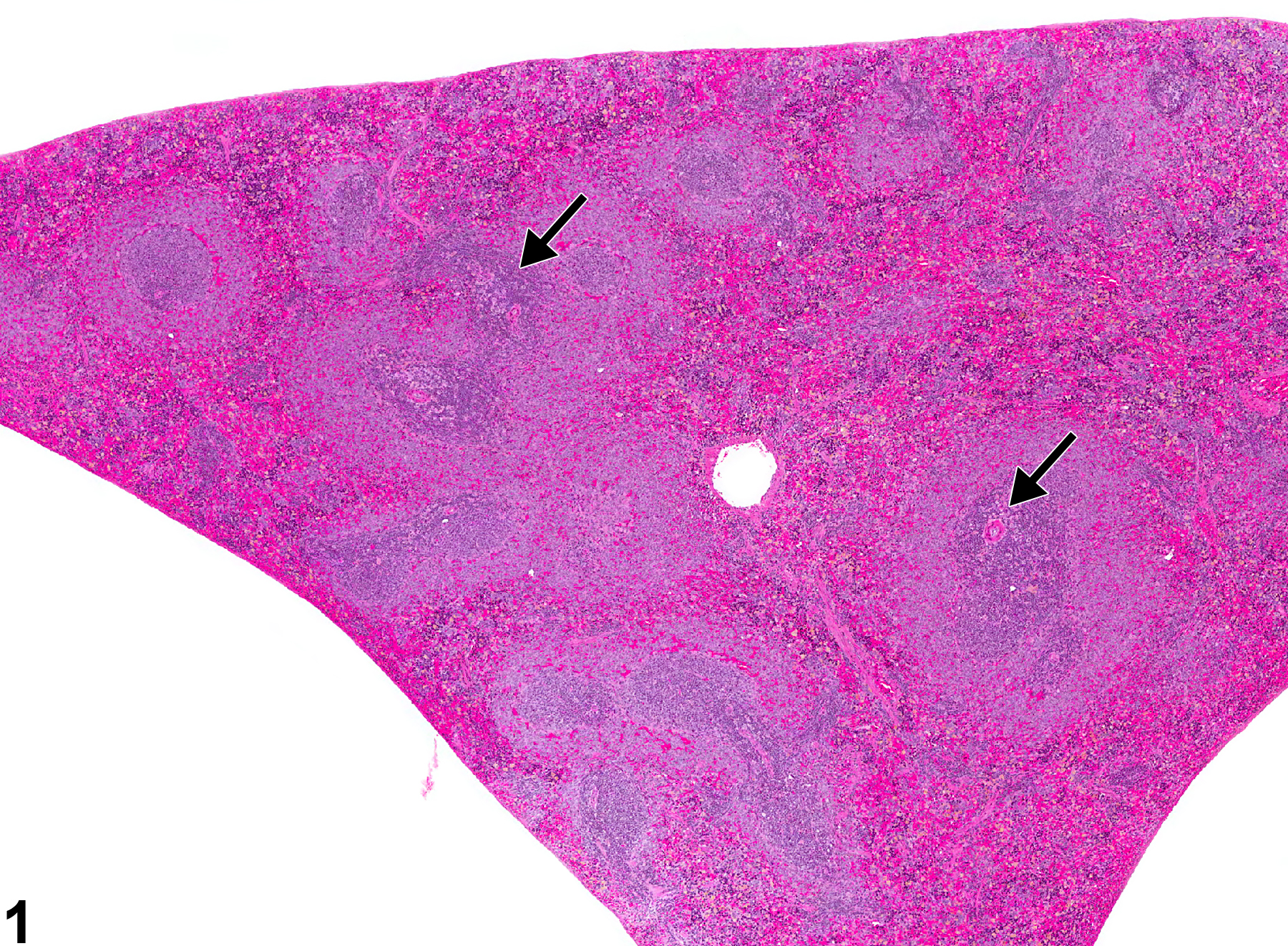
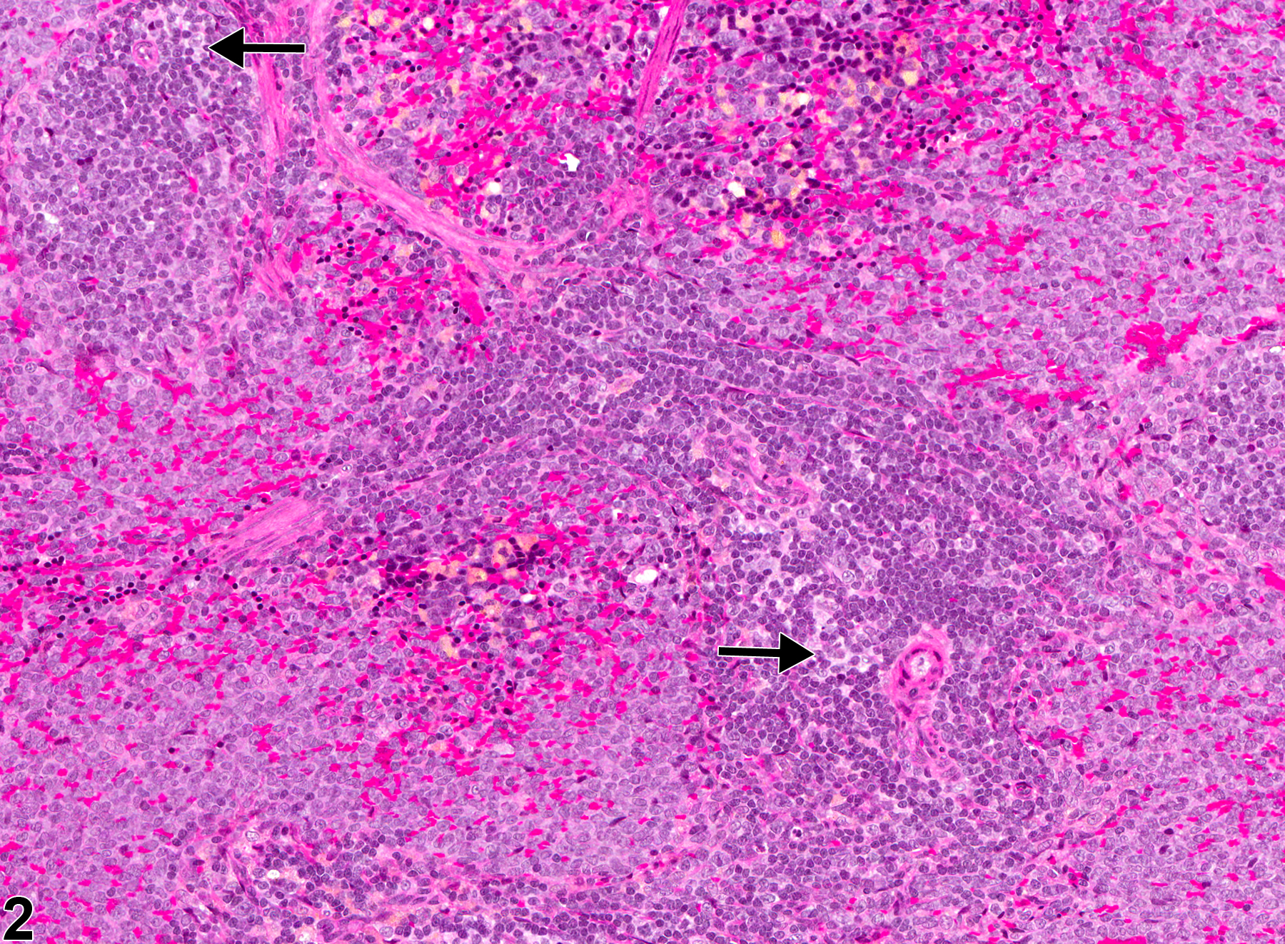
Spleen, White pulp - Atrophy in a female F344/N rat from a subchronic study. Overall area and cellularity of the white pulp (periarteriolar lymphatic sheaths and follicles) are decreased (arrows), although the marginal zones appear to be within normal limits.
All Images

Spleen, White pulp - Atrophy in a female F344/N rat from a subchronic study. Overall area and cellularity of the white pulp (periarteriolar lymphatic sheaths and follicles) are decreased (arrows), although the marginal zones appear to be within normal limits.

Spleen, White pulp - Atrophy in a female F344/N rat from a subchronic study (higher magnification of Figure 1). Decreased cellularity and area of the periarteriolar lymphatic sheath region (arrows) are evident.

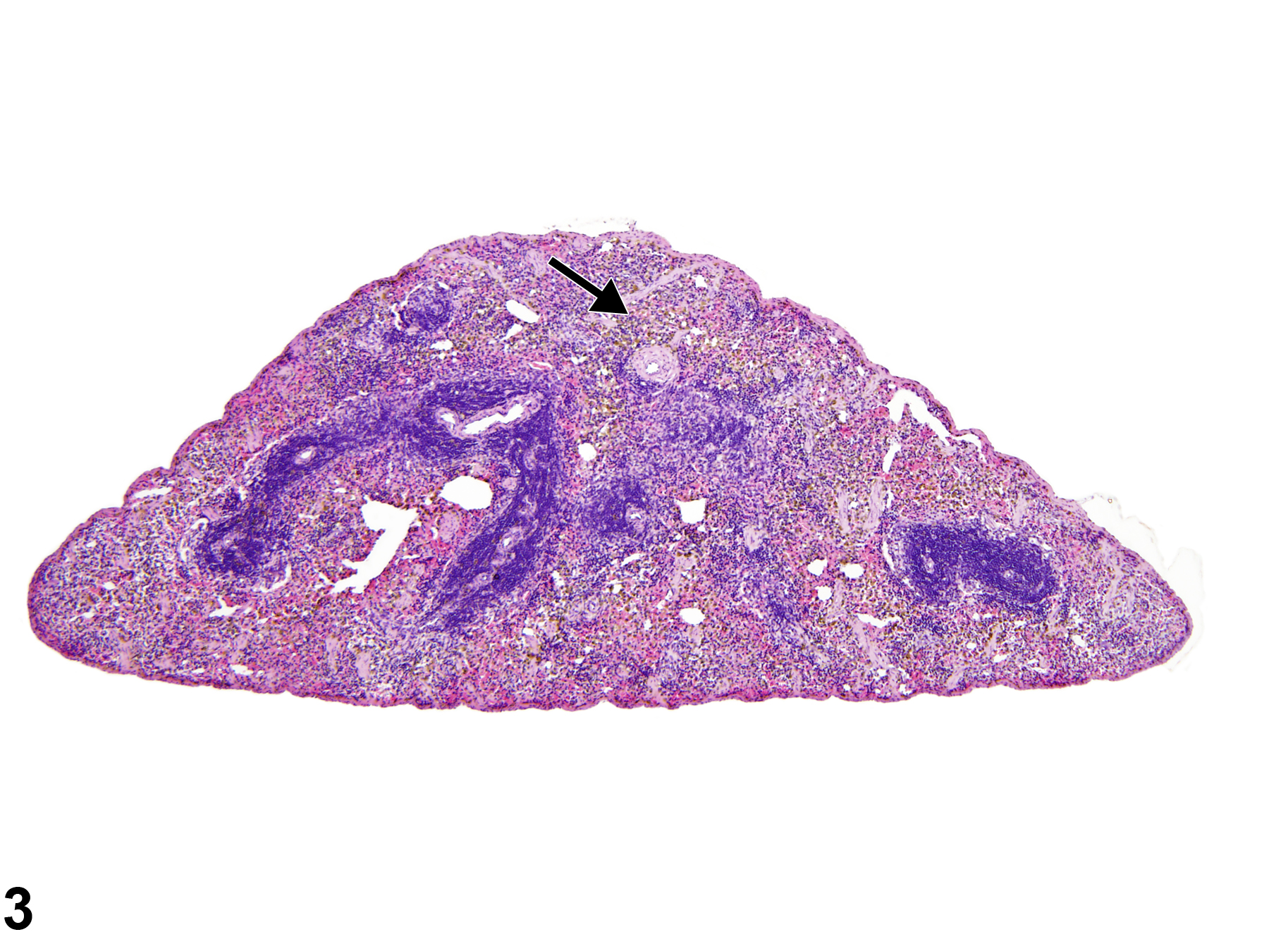
Spleen - Atrophy in a female B6C3F1/N mouse from a subchronic study. Both red pulp (arrow) and white pulp (follicles, periarteriolar lymphatic sheaths, marginal zones) are atrophied.

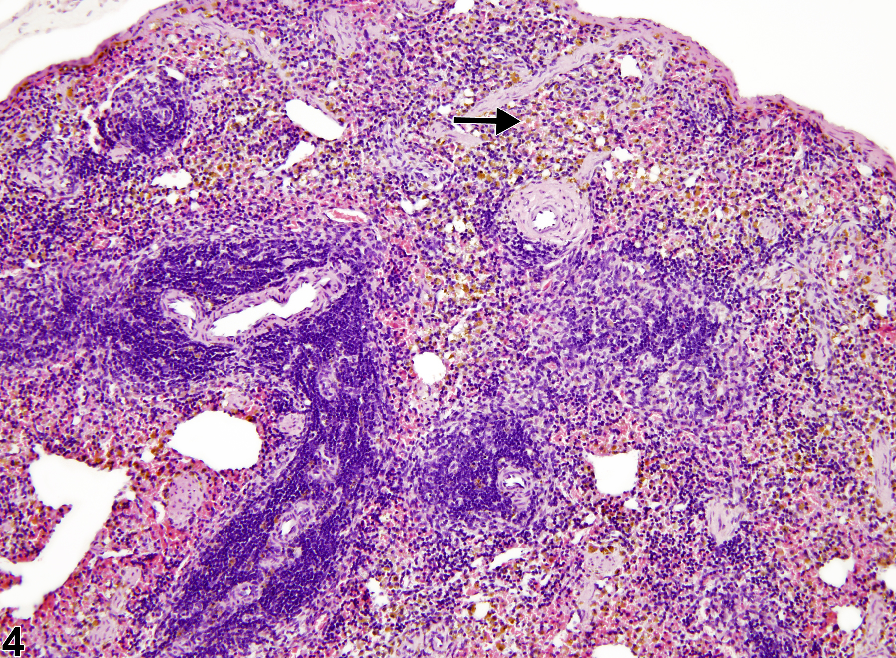
Spleen - Atrophy in a female B6C3F1/N mouse from a subchronic study (higher magnification of Figure 3). In addition to white pulp atrophy, red pulp elements are decreased, and there is a moderate amount of pigment within the red pulp compartment (arrow).