U.S. flag

An official website of the United States government

Dot gov

The .gov means it's official.
Federal government websites often end in .gov or .mil. Before sharing sensitive information, make sure you're on a federal government site.

Https

The site is secure.
The https:// ensures that you are connecting to the official website and that any information you provide is encrypted and transmitted securely.

Skip to Main Navigation
Skip to Page Content
Skip to Atlas Navigation

Spleen - Pigment

Image of pigment in the spleen from a male F344/N rat in a chronic study
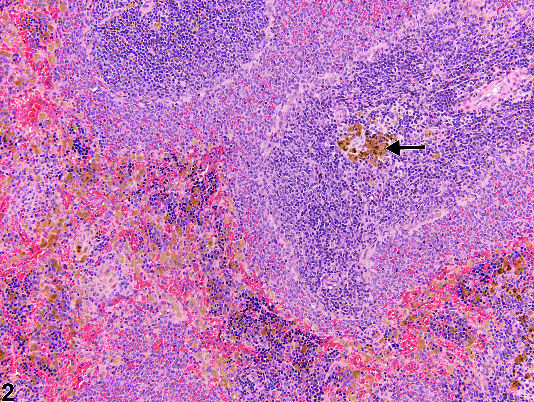
Spleen - Pigment in a male F344/N rat from a chronic study. Multifocal macrophages with intracytoplasmic dark-brown granular pigment (arrow) are present within the splenic red pulp.
Figure 1 of 2
Image of pigment in the spleen from a female Harlan Sprague-Dawley rat in a chronic study
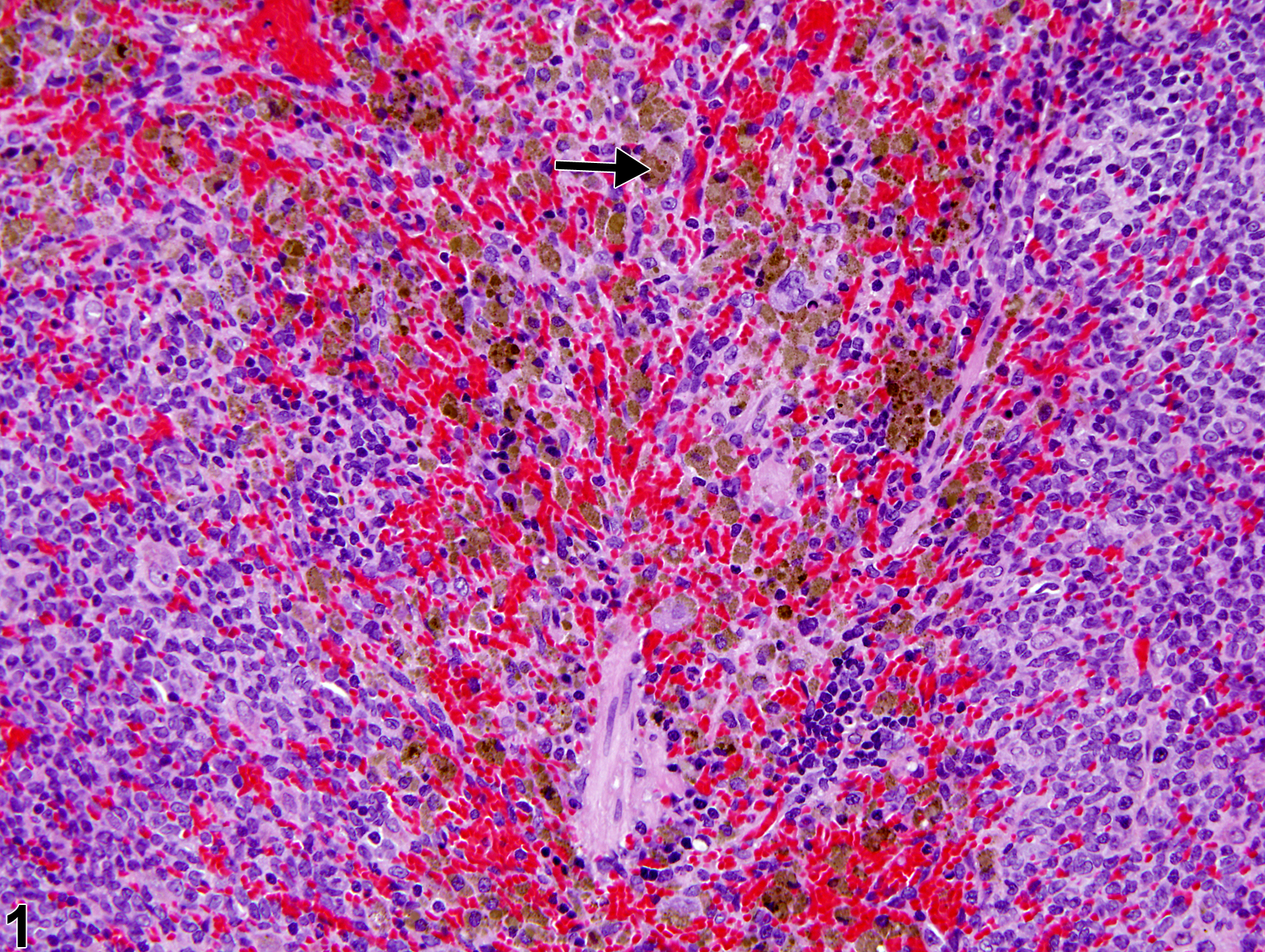
Spleen - Pigment in a female Sprague-Dawley rat from a chronic study. Macrophages with intracytoplasmic golden-brown pigment (arrow) are focally present within the splenic white pulp.
Figure 2 of 2
next arrow

comment:

The presence of a small amount of intracytoplasmic pigment within the splenic red pulp macrophages is a common background finding in rodents. Pigment can be hemosiderin, ceroid/lipofuscin, melanin, or test article related. It is typically found in the red pulp ( Figure 1image opens in a pop-up window , arrow) but may also be present in the marginal zone and/or periarteriolar lymphatic sheaths of the white pulp ( Figure 2image opens in a pop-up window , arrow), the capsule, or trabeculae. Hemosiderin pigment is the result of macrophage engulfment of red blood cells (usually effete or damaged) or free hemoglobin and may be increased in aging rodents and/or such conditions as treatment-induced hemolytic anemia or methemoglobinemia, chronic congestion, increased hematopoietic cell proliferation, and erythrophagocytosis. Hemosiderin deposits may also be observed with focal hemorrhage secondary to malignant neoplasms. The incidence of hemosiderin pigment in aging rodents is usually higher in females than in males for both rats and mice. Ceroid/lipofuscin, derived from oxidation and polymerization of unsaturated lipids, may also be present within splenic macrophages but occurs less commonly than hemosiderin. Hemosiderin can be identified with Perl’s Prussian blue special stain, whereas ceroid/lipofuscin is acid-fast positive. Melanin pigment can be seen in trabecular melanocytes or focally in the red pulp of black mice. Fontana-Masson or Schmorl’s ferric-ferricyanide reduction stain can be used to identify melanin granules. Pigment may be subclassified by type (i.e., hemosiderin, ceroid/ lipofuscin, melanin) only when confirmed by special stains.

recommendation:

Pigment in the red and/or white pulp of the spleen should be diagnosed when increased or decreased due to treatment and assigned a severity grade. A diagnostic modifier (i.e., "increased" or "decreased") should be included in the diagnosis to indicate whether the pigment is increased or decreased relative to concurrent controls. Definitive pigment identification is often difficult in histological sections, even with a battery of special stains. Therefore, a diagnosis of "pigment" (as opposed to diagnosing the type of pigment, e.g., hemosiderin or lipofuscin) is most appropriate. Morphological features of the pigment should be described in the pathology narrative. If the presence of pigment is secondary to age or not toxicologically meaningful, the pigment should not be diagnosed. The pathologist should use his or her judgment in deciding whether secondary deposits of pigment are prominent enough to warrant a separate diagnosis.

references:

National Toxicology Program. 2010. NTP TR-558. Toxicology and Carcinogenesis Studies of 3,3′,4,4′-Tetrachloroazobenzene (TCAB) [CAS No. 14047-09-7] in Harlan Sprague-Dawley Rats and B6C3F1 Mice (Gavage Studies). NTP, Research Triangle Park, NC.
Abstract: https://ntp.niehs.nih.gov/go/33564

National Toxicology Program. 2011. NTP TR-570. Toxicology and Carcinogenesis Studies of α,β-Thujone (CAS No. 76231-76-0) in F344/N Rats and B6C3F1 Mice (Gavage Studies). NTP, Research Triangle Park, NC.
Abstract: https://ntp.niehs.nih.gov/go/36137

Stefanski SA, Elwell MR, Stromberg PC. 1990. Spleen, lymph nodes, and thymus. In: Pathology of the Fischer Rat: Reference and Atlas (Boorman GA, Eustis SL, Elwell MR, Montgomery CA, MacKenzie WF, eds). Academic Press, San Diego, 369-394.

Suttie AW. 2006. Histopathology of the spleen. Toxicol Pathol 34:466-503.
Full Text: http://tpx.sagepub.com/content/34/5/466.full.pdf

Ward JM, Rehg JE, Morse HC III. 2012. Differentiation of rodent immune and hematopoietic system reactive lesions from neoplasias. Toxicol Pathol 40:425-434.
Abstract: https://www.ncbi.nlm.nih.gov/pubmed/22215512

Ward JM, Mann PC, Morishima H, Frith CH. 1999. Thymus, spleen, and lymph nodes. In: Pathology of the Mouse (Maronpot RR, ed). Cache River Press, Vienna, IL, 333-360.