

Integumentary System
Skin - Amyloid
Narrative
Aigelsreiter A, Janig E, Stumptner C, Fuchsbichler A, Zatloukal K, Denk H. 2007. How a cell deals with abnormal proteins. Pathogenetic mechanisms in protein aggregation diseases. Pathobiology 74:145-158.
Abstract: https://www.ncbi.nlm.nih.gov/pubmed/17643060Greaves P. 2007. Integumentary System. In: Histopathology of Preclinical Toxicity Studies. Interpretation and Relevance in Drug Safety Evaluation, 3rd ed. Elsevier, Amsterdam.
Abstract: http://www.sciencedirect.com/science/book/9780444527714Peckham JC, Heider K. 1999. Skin and subcutis. In: Pathology of the Mouse: Reference and Atlas (Maronpot RR, Boorman GA, Gaul BW, eds). Cache River Press, Vienna, IL, 555-612.
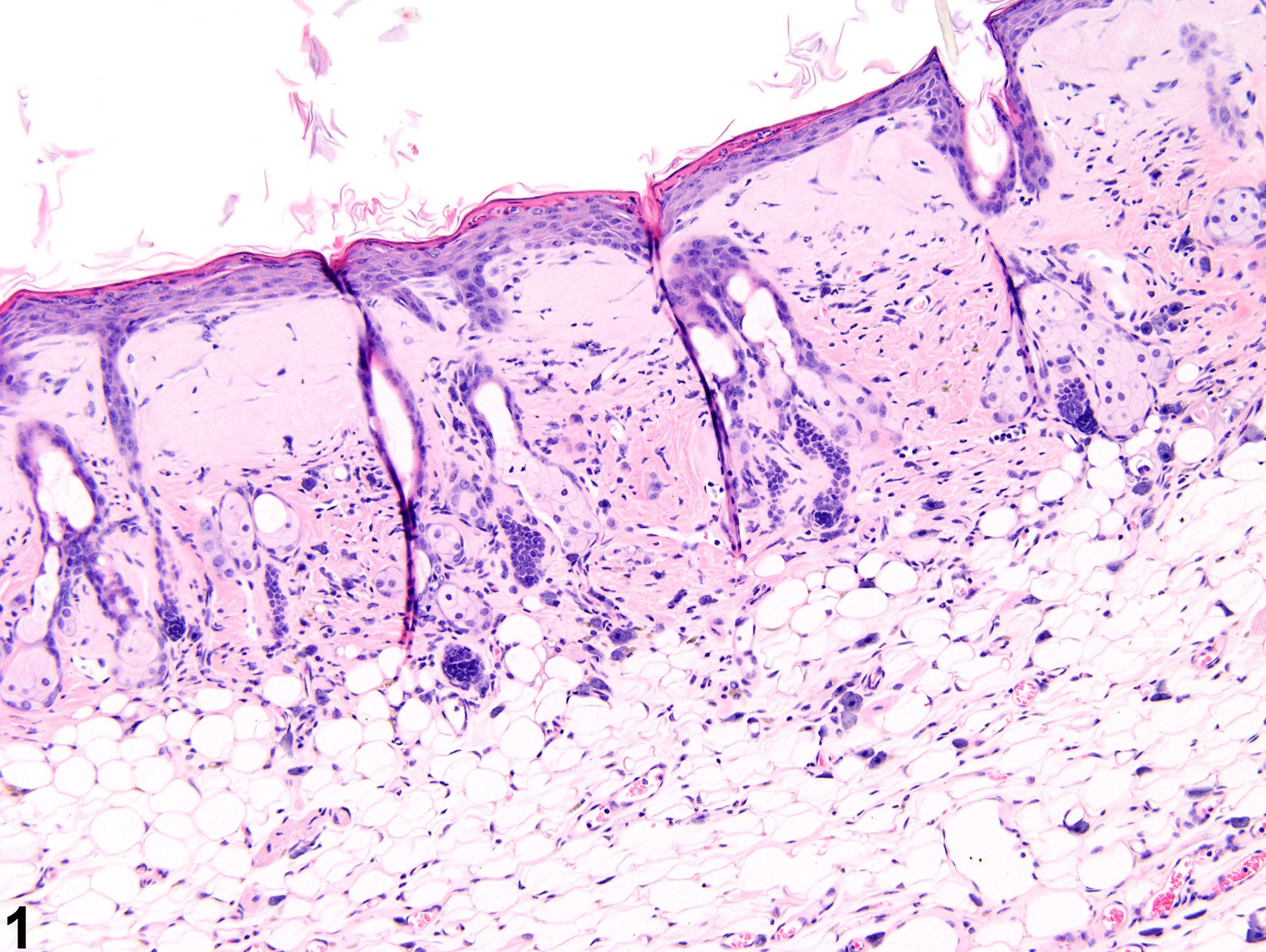
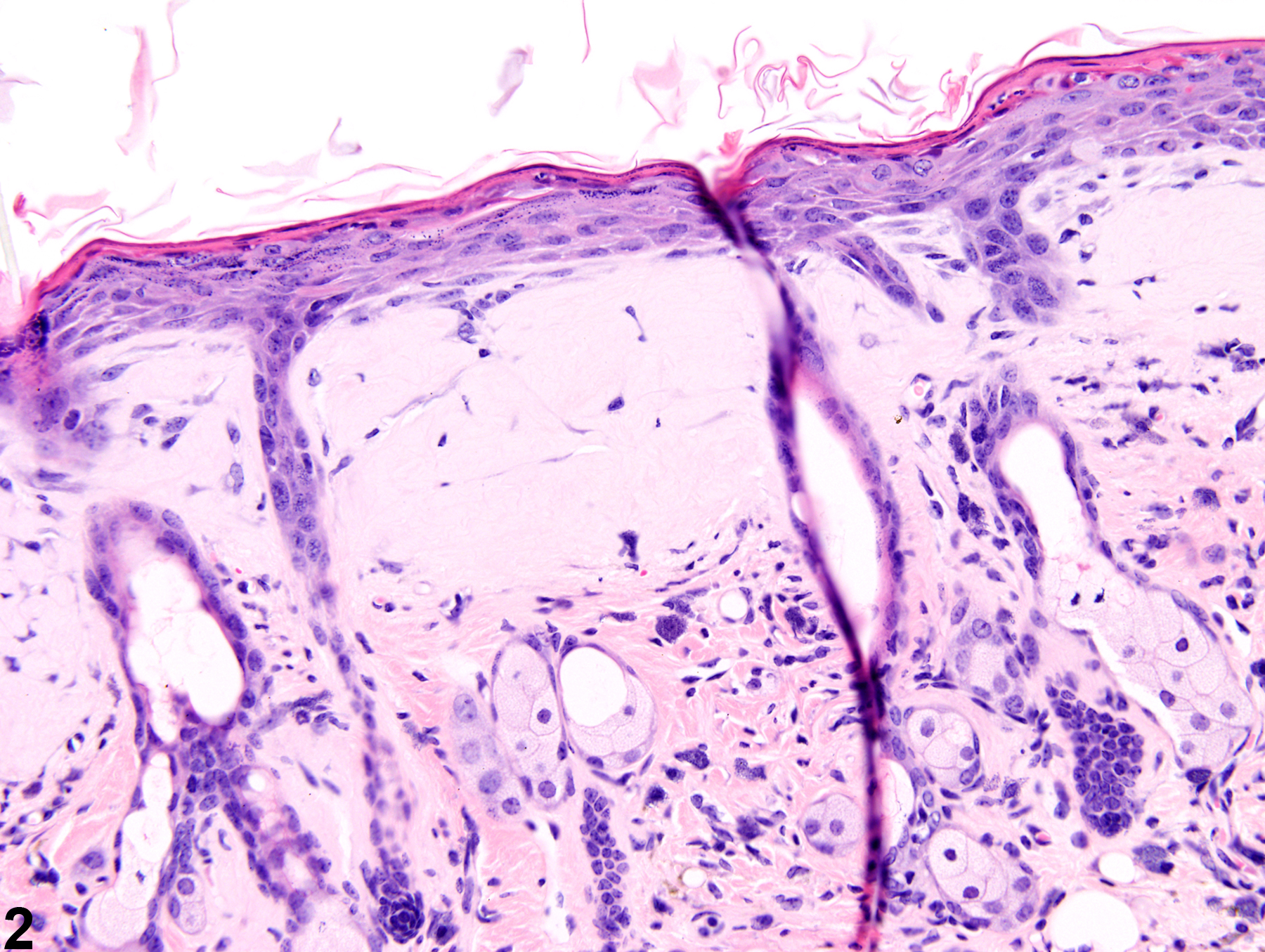
Amyloid-accumulations of amorphous, eosinophilic, extracellular material in a female Swiss CD-1 mouse from a chronic study.
All Images

Amyloid-accumulations of amorphous, eosinophilic, extracellular material in a female Swiss CD-1 mouse from a chronic study.

Amyloid-accumulations of amorphous, eosinophilic, extracellular material in a female Swiss CD-1 mouse from a chronic study.