

Reproductive System, Male
Coagulating Gland - Atrophy
Narrative
Boorman GA, Elwell MR, Mitsumori K. 1990. Male accessory sex glands, penis, and scrotum. In: Pathology of the Fischer Rat: Reference and Atlas (Boorman GA, Eustis SL, Elwell MR, Montgomery CA, MacKenzie WF, eds). Academic Press, San Diego, 419-428.
Abstract: http://www.ncbi.nlm.nih.gov/nlmcatalog/9002563Bosland MC, Tuomari DL, Elwell MR, Shirai T, Ward JM, McConnell RF. 1998. Proliferative lesions of the prostate and other accessory sex glands in male rats, URG-4. In: Guidelines for Toxicologic Pathology. STP/ARP/AFIP, Washington, DC, 1-20.
Markus RP, Goncalo MC, Lapa AJ. 1981. Castration atrophy and pharmacological reactivity of the rat coagulating gland. Braz J Med Res 14:181-185.
Abstract: http://www.ncbi.nlm.nih.gov/pubmed/6121593Suwa T, Nyska A, Haseman JK, Mahler JF, Maronpot RR. 2002. Spontaneous lesions in control B6C3F1 mice and recommended sectioning of male accessory sex organs. Toxicol Pathol 30(2):228- 234.
Abstract: http://www.ncbi.nlm.nih.gov/pubmed/11950166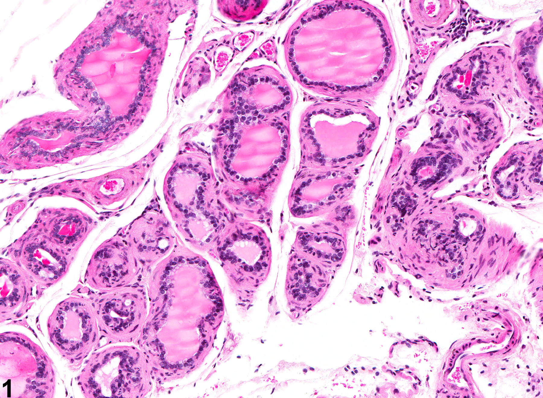
Coagulating Gland - Atrophy. Atrophy of the coagulating gland in a male B6C3F1 mouse from a chronic study.
All Images

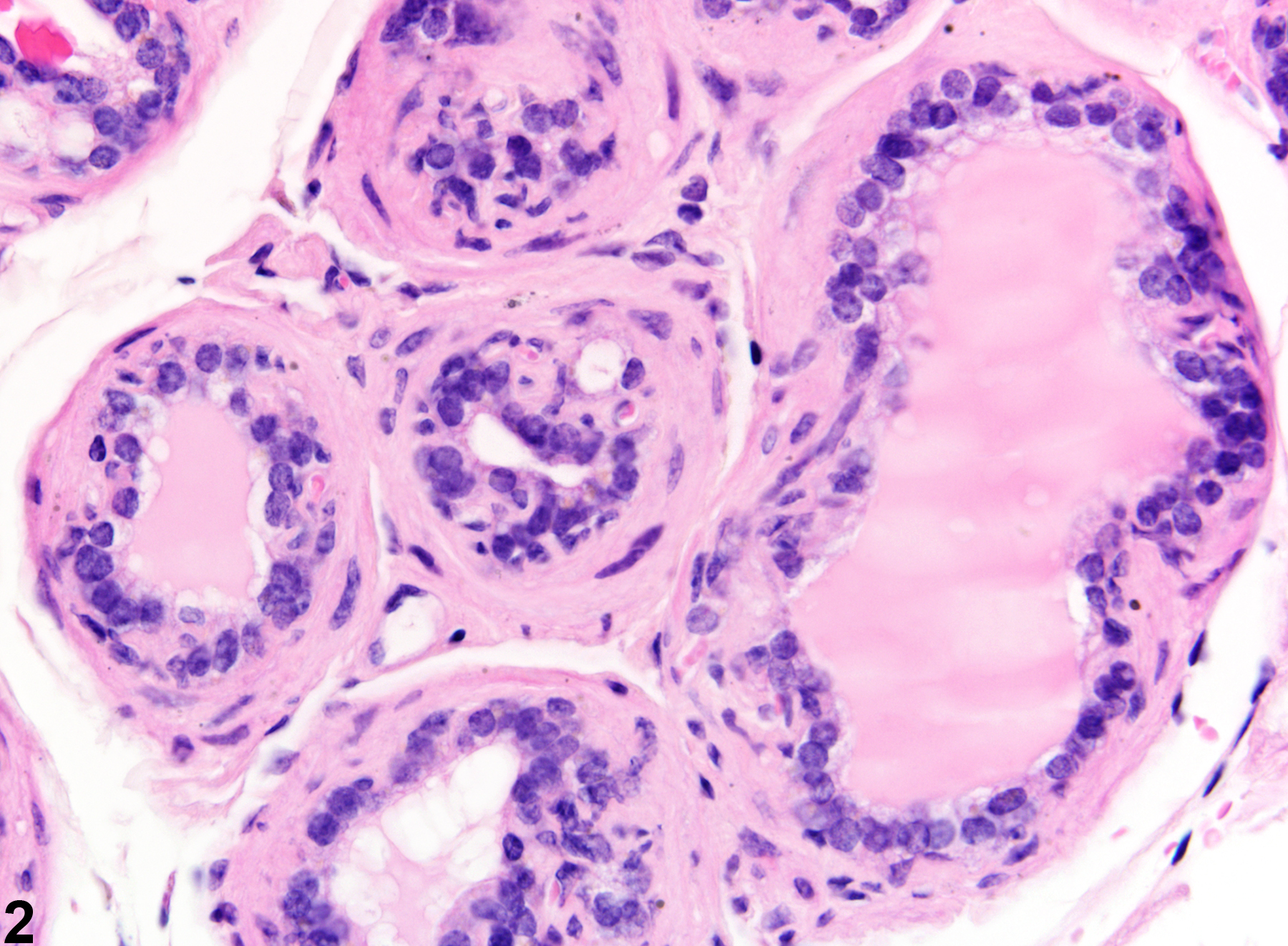
Coagulating Gland - Atrophy. Atrophy of the coagulating gland in a male B6C3F1 mouse from a chronic study.
Coagulating Gland - Atrophy. Atrophy of the coagulating gland in a male B6C3F1 mouse from a chronic study.