

Reproductive System, Male
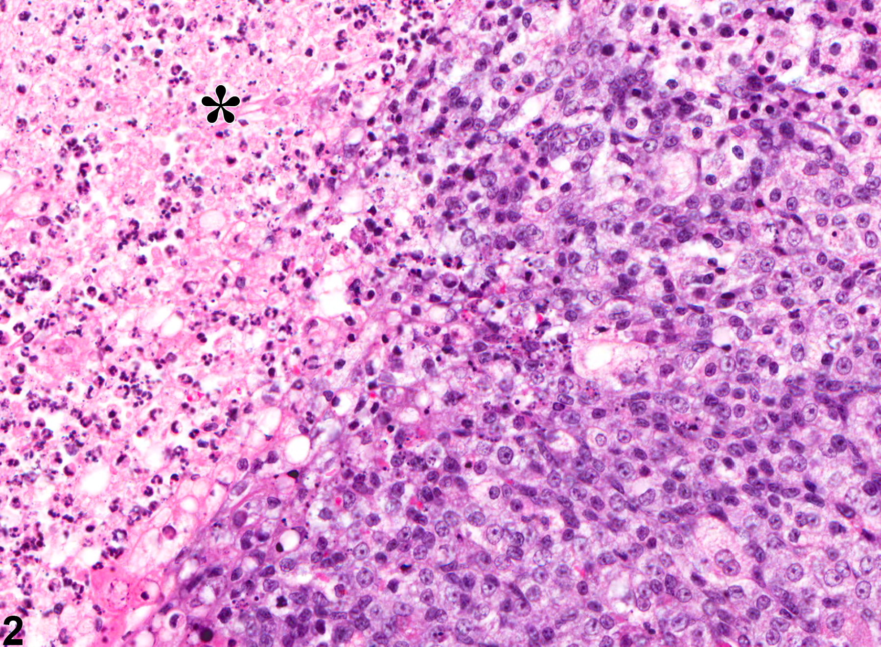
Preputial Gland - Necrosis
Narrative
Boorman GA, Elwell MR, Mitsumori K. 1990. Male accessory sex glands, penis, and scrotum. In: Pathology of the Fischer Rat: Reference and Atlas (Boorman GA, Eustis SL, Elwell MR, Montgomery CA, MacKenzie WF, eds). Academic Press, San Diego, 419-428.
Abstract: http://www.ncbi.nlm.nih.gov/nlmcatalog/9002563Gordon LR, Majka JA, Boorman GA. 1996. Spontaneous nonneoplastic and neoplastic lesions and experimentally induced neoplasms of the testes and accessory sex glands. In: Pathobiology of the Aging Mouse, Vol 1 (Mohr U, Dungworth DL, Capen CC, Carlton WW, Sundberg JP, Ward JM, eds). ILSI Press, Washington, DC, 421-441.
Abstract: http://catalog.hathitrust.org/Record/008994685Haines DC, Eustis SL. 1990. Specialized sebaceous glands. In: Pathology of the Fischer Rat: Reference and Atlas (Boorman GA, Eustis SL, Elwell MR, Montgomery CA, MacKenzie WF, eds). Academic Press, San Diego, 279-293.
Abstract: http://www.ncbi.nlm.nih.gov/nlmcatalog/9002563Rudmann D, Cardiff R, Chouinard L, Goodman D, Kuttler K, Marxfeld H, Molinolo A, Treumann S, Yoshizawa K. 2012. Proliferative and nonproliferative lesions of the rat and mouse mammary, Zymbal's, preputial, and clitoral glands. Toxicol Pathol 40:7S-39S.
Abstract: http://www.ncbi.nlm.nih.gov/pubmed/22949413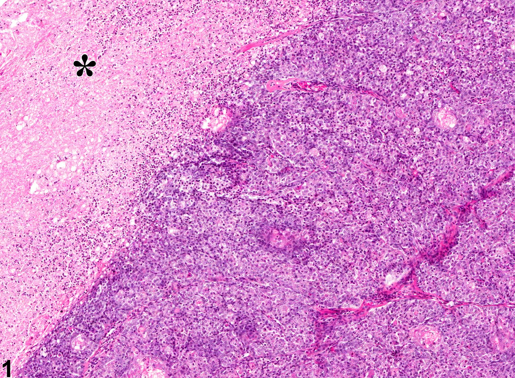
Preputial Gland - Necrosis. Asterisk indicates area of necrosis from a male F344/N rat in a chronic study.
All Images

Preputial Gland - Necrosis. Asterisk indicates area of necrosis from a male F344/N rat in a chronic study.

Preputial Gland - Necrosis. Higher magnification of Figure 1. Asterisk indicates accumulation of cellular, pyknotic, and karyorrhectic debris from a male F344/N rat in a chronic study.