

Reproductive System, Male
Seminal Vesicle - Dilation
Narrative
Radovsky A, Mitsumori K, Chapin RE. 1999. Male reproductive tract. In: Pathology of the Mouse: Reference and Atlas (Maronpot RR, Boorman GA, Gaul BW, eds). Cache River Press, Vienna, IL, 381-407.
Suwa T, Nyska A, Peckham JC, Hailey JR, Mahler JF, Haseman JK, Maronpot RR. 2001. A retrospective analysis of background lesions and tissue accountability for male accessory sex organs in Fischer-344 rats. Toxicol Pathol 29(4):467-478.
Abstract: http://www.ncbi.nlm.nih.gov/pubmed/11560252Suwa T, Nyska A, Haseman JK, Mahler JF, Maronpot RR. 2002. Spontaneous lesions in control B6C3F1 mice and recommended sectioning of male accessory sex organs. Toxicol Pathol 30(2):228-234.
Abstract: http://www.ncbi.nlm.nih.gov/pubmed/11950166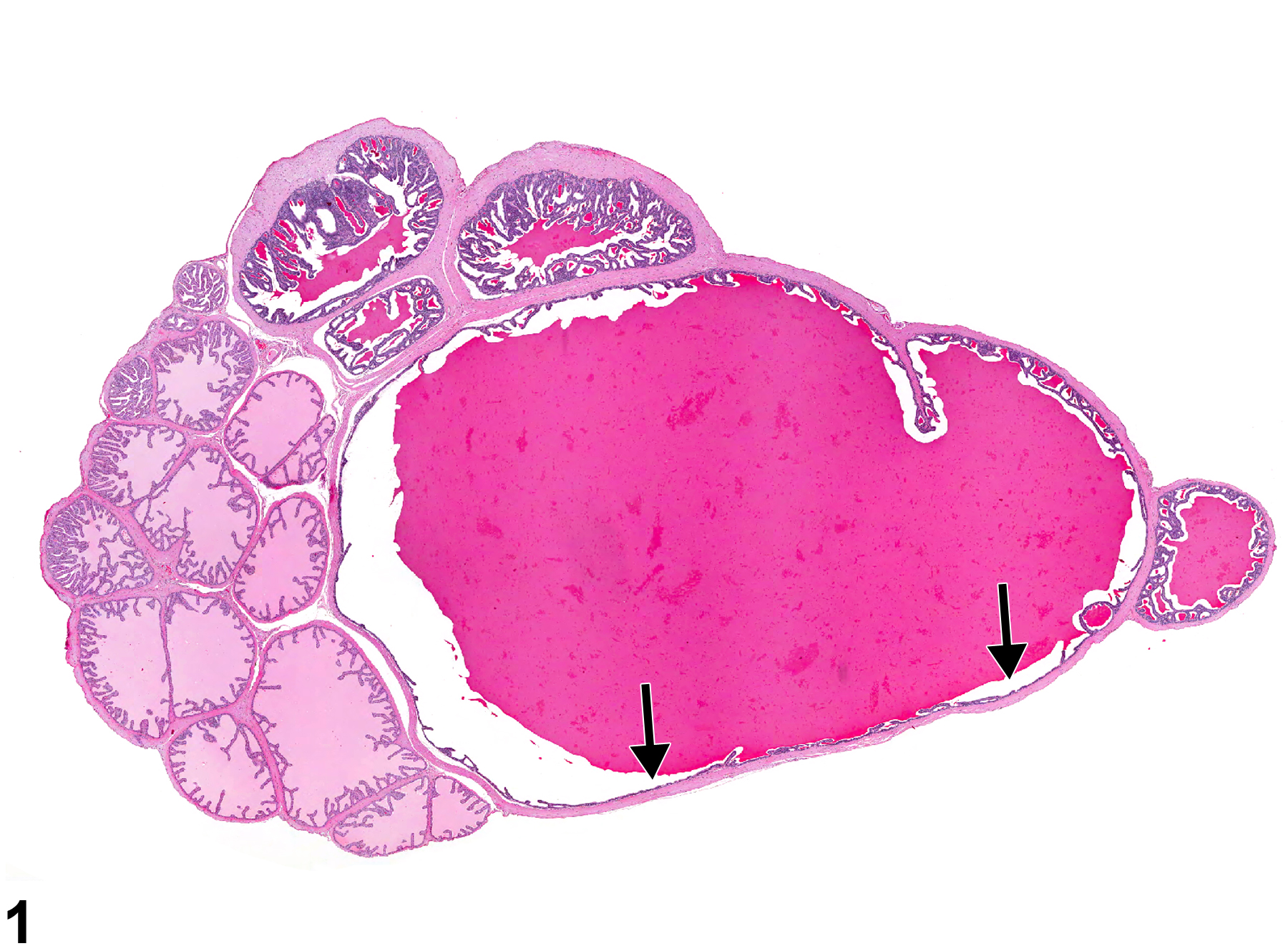
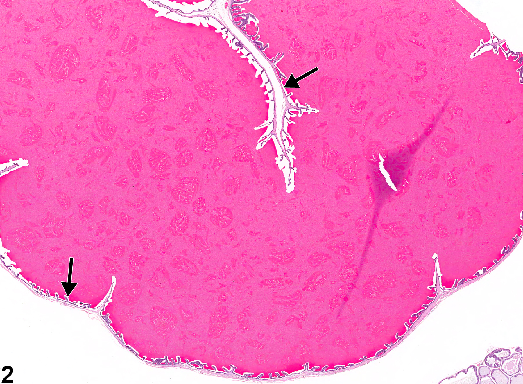
Seminal Vesicle - Dilation. Arrows indicate flattened epithelium in the distended acinus in a male F344/N rat from a chronic study.
All Images

Seminal Vesicle - Dilation. Arrows indicate flattened epithelium in the distended acinus in a male F344/N rat from a chronic study.

Seminal Vesicle - Dilation. Arrows indicate flattened epithelium in the distended acinus in a male F344/N rat from a chronic study.