

Nervous System
Brain, Neuron - Degeneration
Narrative
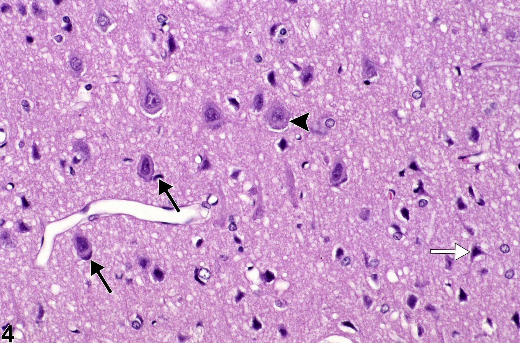
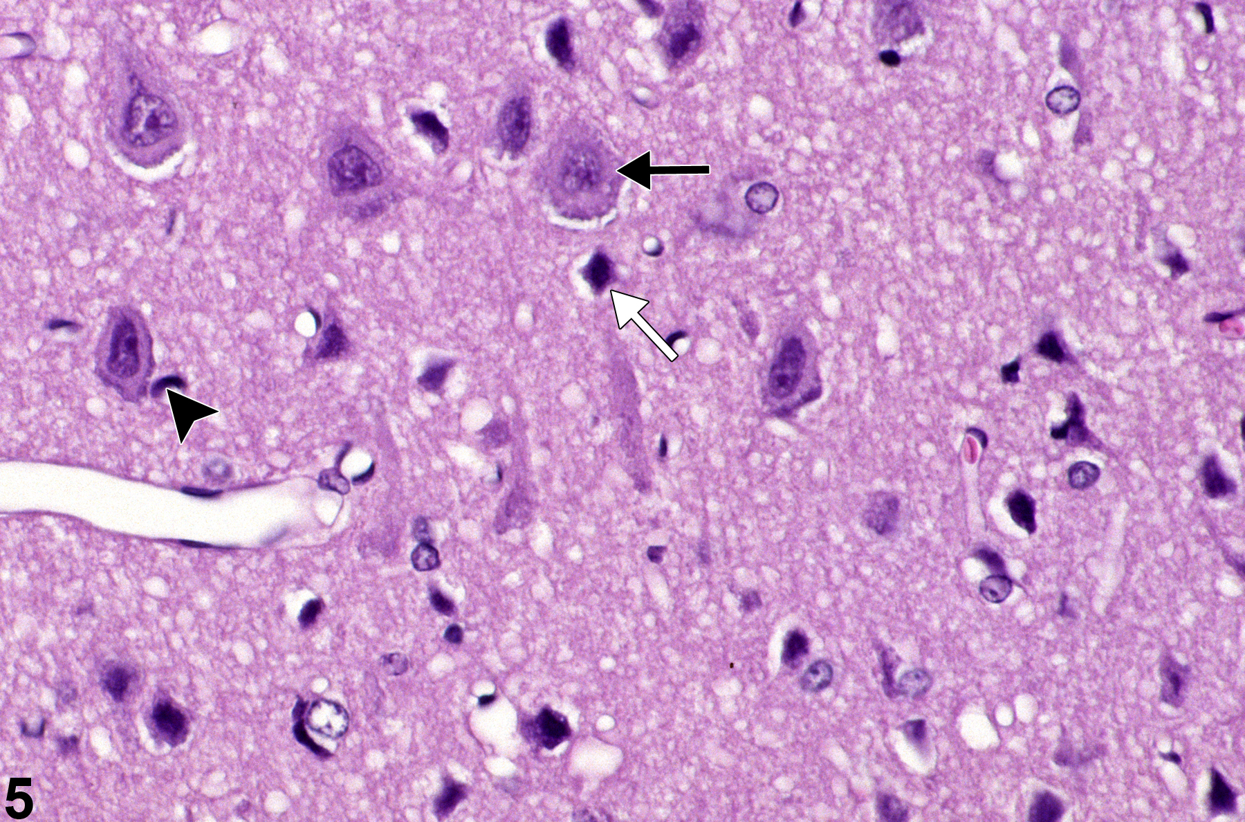
Figure 4 and Figure 5 depict a commonly overlooked subtle and early form of neuronal injury known as “granulovacuolar change.” The perikaryon is slightly swollen and contains small vacuoles and granularity (arrowheads), suggesting membrane injury and distension of the cytoplasmic organelles such as the endoplasmic reticulum and mitochondria. In these images, it is accompanied by other ancillary evidence of neuronal injury, such as central chromatolysis (black arrows) and genuine basophilic “chronic” nerve cell change (white arrows). Various stages of neuronal and astrocytic degeneration as documented by light and electron microscopy are available in a sequenced study by Cavanagh et al. (1998) of the effects of a mycotoxin on the rat cerebellum.
Cavanagh JB, Holton JL, Nolan CC, Ray DE, Naik JT, Mantle PG. 1998. The effects of the tremorgenic mycotoxin penitrem A on the rat cerebellum. Vet Pathol 35:53-63.
Abstract: http://www.ncbi.nlm.nih.gov/pubmed/9545135Colman JR, Nowocin KJ, Switzer RC, Trusk TC, Ramsdell JS. 2005. Mapping and reconstruction of domoic acid-induced neurodegeneration in the mouse brain. Neurotoxicol Teratol 27:753-767.
Abstract: http://www.ncbi.nlm.nih.gov/pubmed/16109471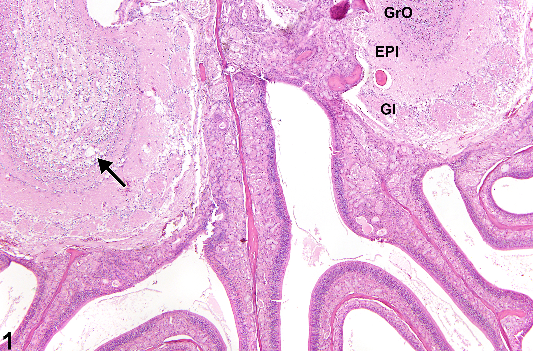
Olfactory bulb neuronal degeneration in a female B6C3F1 mouse from a chronic study. The arrow indicates the vacuolation and neuronal degeneration of the olfactory bulb. Gl, glomerular layer; EPl, external plexiform layer; GrO, internal granule cell layer.
All Images

Olfactory bulb neuronal degeneration in a female B6C3F1 mouse from a chronic study. The arrow indicates the vacuolation and neuronal degeneration of the olfactory bulb. Gl, glomerular layer; EPl, external plexiform layer; GrO, internal granule cell layer.

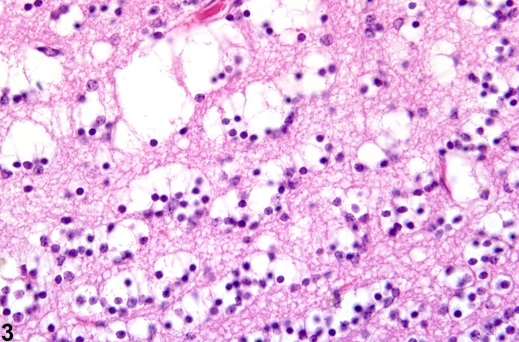
Higher magnification of the internal granule cell layer in the normal olfactory bulb in a female B6C3F1 mouse from a chronic study.

The contralateral side shows the prominent vacuolation and neuronal degeneration.

Granulovacuolar neuronal degeneration in a male F344/N rat from an acute study. The black arrowhead indicates neuronal granulovacuolar change, the black arrows identify neurons with central chromatolysis, and the white arrow identifies a genuine basophilic necrotic neuron.

Granulovacuolar neuronal degeneration in a male F344/N rat from an acute study. Higher magnification of Figure 4. The black arrow indicates neuronal granulovacuolar change, the arrowhead identifies a neuron with central chromatolysis, and the white arrow identifies a genuine basophilic necrotic neuron.