

Respiratory System
Nose, Epithelium - Accumulation, Hyaline Droplet
Narrative
Boorman GA, Morgan KT, Uraih LC. 1990. Nose, larynx, and trachea. In: Pathology of the Fischer Rat: Reference and Atlas (Boorman GA, Eustis SL, Elwell MR, eds). Academic Press, San Diego, 315-337.
Herbert RA, Leninger JR. 1999. Nose, larynx, and trachea. In: Pathology of the Mouse: Reference and Atlas (Maronpot RR, ed). Cache River Press, Vienna, IL, 259-292.
Monticello TM, Morgan KT, Uraih LC. 1990. Nonneoplastic nasal lesions in rats and mice. Environ Health Perspect 85:249-274.
Full Text: https://www.ncbi.nlm.nih.gov/pmc/articles/PMC1568333/Renne R, Brix A, Harkema J, Kittel B, Lewis D, March T, Nagano K, Pino M, Rittinghausen S, Rosenbruch M, Tellier P, Wohrmann T. 2009. Proliferative and nonproliferative lesions of the rat and mouse respiratory tract. Toxicol Pathol 37(7 suppl):5S-73S.
Abstract: https://www.ncbi.nlm.nih.gov/pubmed/20032296St Clair MGB, Morgan KT. 1992. Changes in the upper respiratory tract. In: Pathology of the Aging Rat (Mohr U, Dungworth DL, Capen CC, eds). International Life Sciences Institute Press, Washington, DC, 111-127.
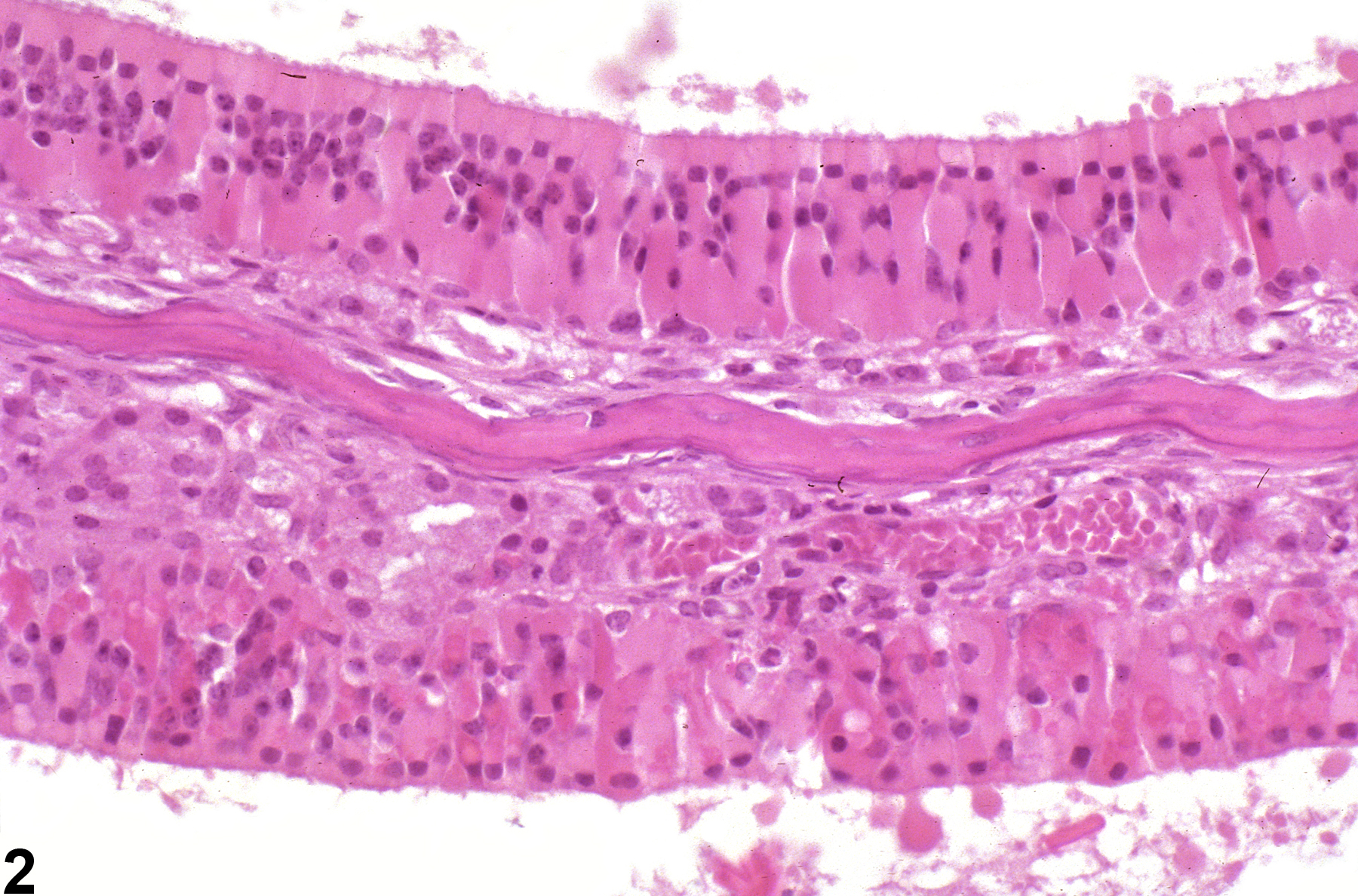
Nose, Respiratory epithelium - Accumulation, Hyaline droplet in a female B6C3F1/N mouse from a chronic study. The respiratory epithelium and cells of the mucosal gland contain eosinophilic globular material.
All Images

Nose, Respiratory epithelium - Accumulation, Hyaline droplet in a female B6C3F1/N mouse from a chronic study. The respiratory epithelium and cells of the mucosal gland contain eosinophilic globular material.

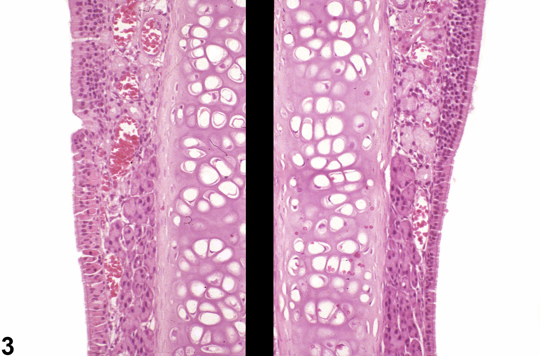
Nose, Olfactory epithelium - Accumulation, Hyaline droplet in a female B6C3F1/N mouse from a chronic study. The turbinate epithelial cells contain homogeneous hyaline material. Image provided courtesy of Dr. R. Miller.

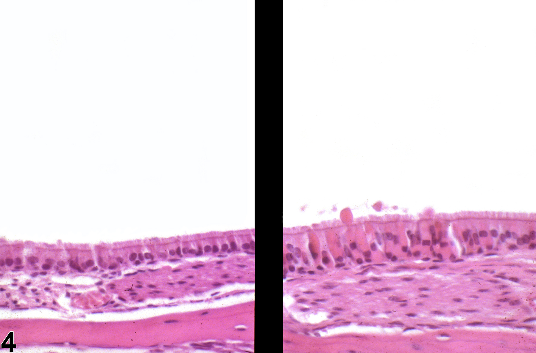
Nose, Respiratory epithelium - Accumulation, Hyaline droplet in a female B6C3F1/N mouse from a subchronic study (left) and normal nasal respiratory epithelium from a control animal (right). Minimal accumulation of hyaline droplet material is present respiratory epithelium at the respiratory and olfactory epithelial junction of the septum in the treated animal (left), but not in the control mouse (right). Image provided courtesy of Dr. R. Miller.

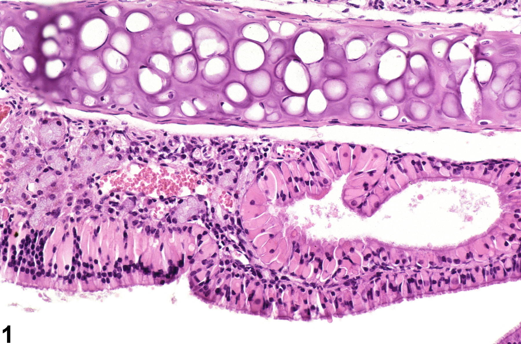
Nose, Respiratory epithelium - Accumulation, Hyaline droplet in a treated female B6C3F1/N mouse (right) and a normal control female B6C3F1/N mouse from a subchronic study (left). Hyaline droplet material is present in the respiratory epithelial cells lining the nasopharyngeal duct from the treated mouse (right) but not in the control mouse (left). Image provided courtesy of Dr. R. Miller.