U.S. flag

An official website of the United States government

Dot gov

The .gov means it's official.
Federal government websites often end in .gov or .mil. Before sharing sensitive information, make sure you're on a federal government site.

Https

The site is secure.
The https:// ensures that you are connecting to the official website and that any information you provide is encrypted and transmitted securely.

Skip to Main Navigation
Skip to Page Content
Skip to Atlas Navigation

Nose, Respiratory Epithelium, Glands - Dilation

Image of dilation in the nose, respiratory epithelium, glands from a male B6C3F1/N mouse in a chronic study
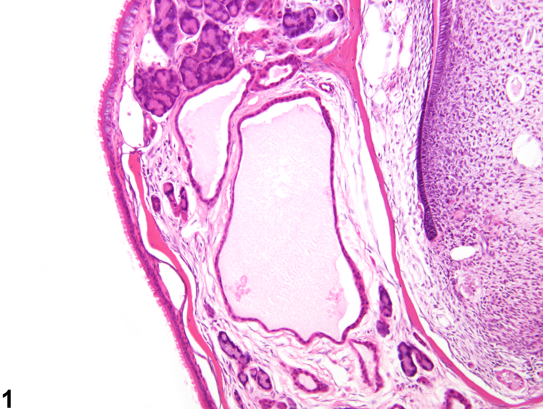
Nose, Respiratory epithelium, Glands - Dilation in a male B6C3F1/N mouse from a chronic study. The glands in the respiratory mucosa are dilated and contain pale eosinophilic material.
Figure 1 of 3
Image of dilation in the nose, respiratory epithelium, glands from a male B6C3F1/N mouse in a chronic study
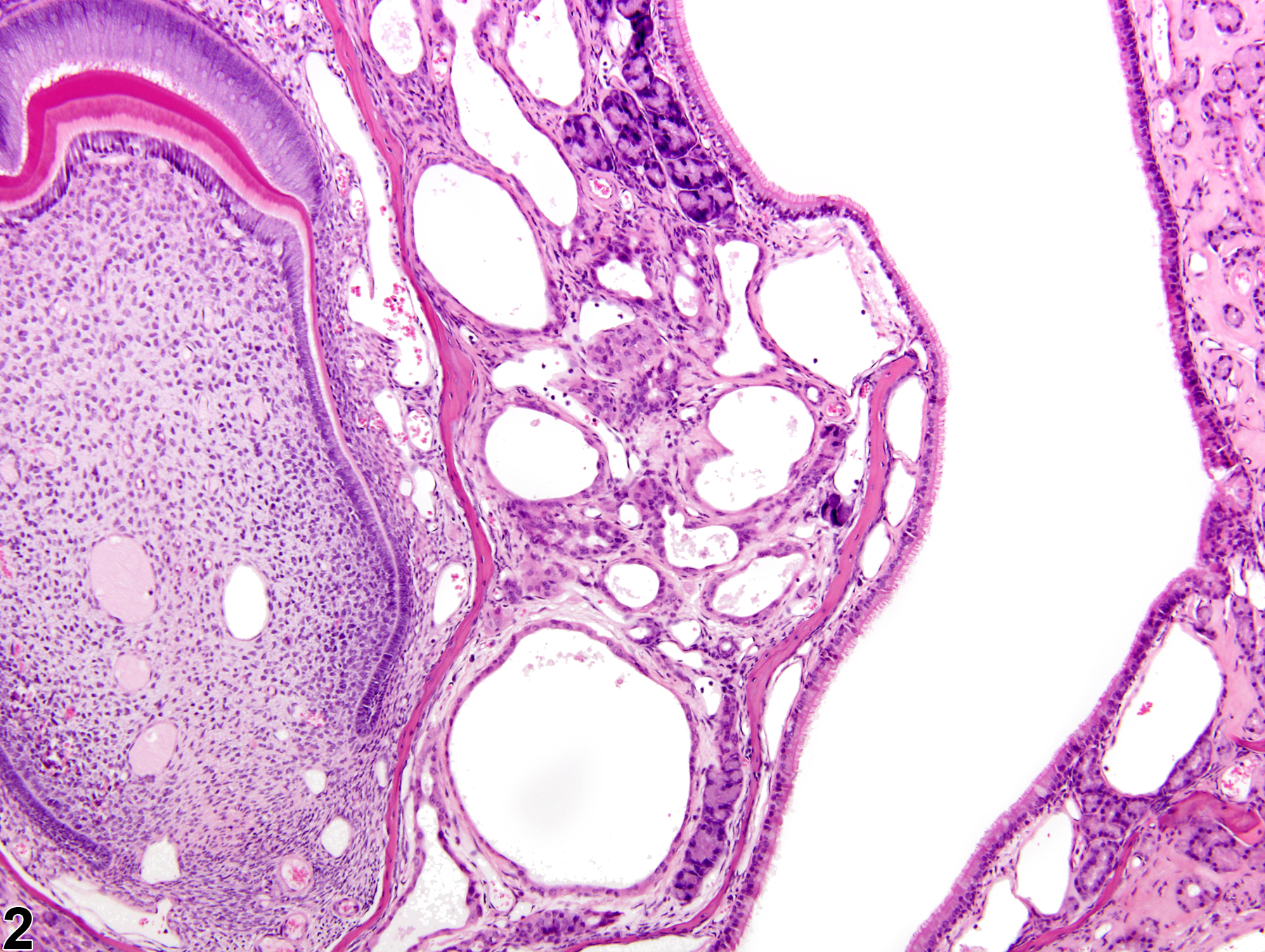
Nose, Respiratory epithelium, Glands - Dilation in a male B6C3F1/N mouse from a chronic study. Variably sized dilated glands are present in the lamina propria of the anterior nasal cavity.
Figure 2 of 3
Image of dilation in the nose, respiratory epithelium, glands from a female B6C3F1/N mouse in a chronic study
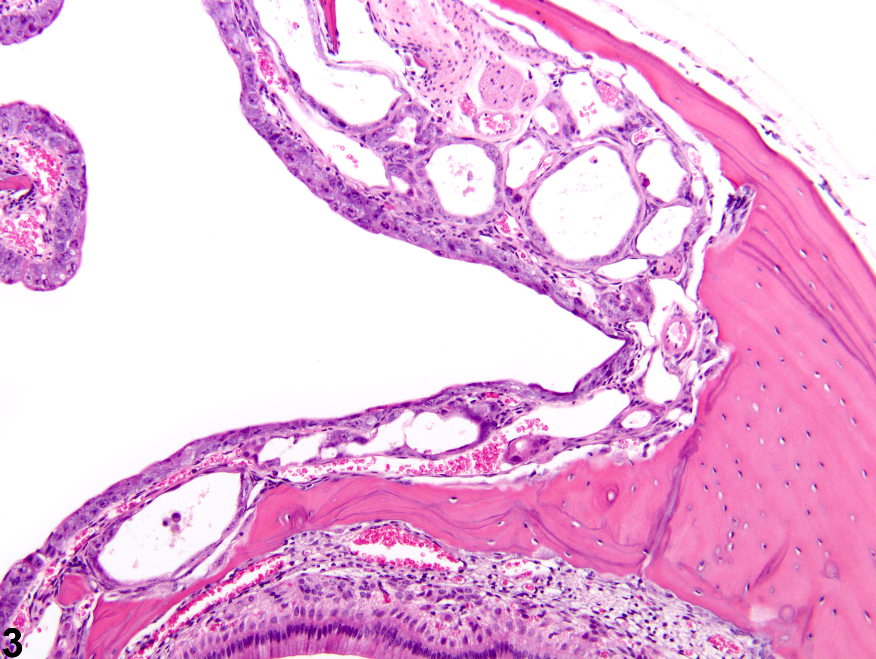
Nose, Respiratory epithelium, Glands - Dilation in a female B6C3F1/N mouse from a chronic study. Multiple dilated glands are present in the lamina propria.
Figure 3 of 3
next arrow

comment:

Dilation of glands in the lamina propria of the respiratory epithelium in excess of normal may alter the normal contour of the nasal septum or turbinates ( Figure 1image opens in a pop-up window , Figure 2image opens in a pop-up window , and Figure 3image opens in a pop-up window ). Glandular dilation typically occurs concurrently with other lesions, such as inflammation and epithelial hyperplasia or metaplasia, but it may be seen separately as a primary change.

recommendation:

"Nose, Respiratory epithelium, Glands - Dilation" should be diagnosed when there is a clear dilation of these glands in the absence of obvious glandular hyperplasia. When diagnosed, this lesion should be assigned a severity grade. If glandular hyperplasia is present concurrently, the hyperplasia should be diagnosed and the dilation described in the narrative. Other lesions, such as inflammation or epithelial degeneration, should be diagnosed separately, if warranted by their prominence or degree of severity.

references:

Boorman GA, Morgan KT, Uraih LC. 1990. Nose, larynx, and trachea. In: Pathology of the Fischer Rat: Reference and Atlas (Boorman GA, Eustis SL, Elwell MR, eds). Academic Press, San Diego, 315-337.

Herbert RA, Leninger JR. 1999. Nose, larynx, and trachea. In: Pathology of the Mouse: Reference and Atlas (Maronpot RR, ed). Cache River Press, Vienna, IL, 259-292.