

Special Senses System
Eye - Phthisis Bulbi
Narrative
Frame SR, Slone TW. 1966. Nonneoplastic and neoplastic changes in the eye. In: Pathobiology of the Aging Mouse, Vol 2 (Mohr U, Dungworth DL, Capen CC, Carlton WW, Sundberg JP, Ward JM, eds). ILSI Press, Washington, DC, 97-103.
Geiss V, Yoshitomi K. 1991. Eyes. In: Pathology of the Mouse: Reference and Atlas (Maronpot RR, Boorman GA, Gaul BW, eds). Cache River Press, Vienna, IL, 471-489.
Hazlett LD, Rosen DD, Berk RS. 1978. Age-related susceptibility to Pseudomonas aeruginosa ocular infections in mice. Infect Immun 20:25-29.
Full Text: http://iai.asm.org/content/20/1/25.full.pdfNewkirk KM, Chandler HL, Parent AE, Young DC, Colitz CMH, Wilkie DA, Kusewitt DF. 2007. Ultraviolet radiation-induced corneal degeneration in 129 mice. Toxicol Pathol 35:817-824.
Full Text: http://tpx.sagepub.com/content/35/6/817.fullNational Toxicology Program. 1979. NTP TR-189. Bioassay of p-Chloroaniline for Possible Carcinogenicity (CAS No. 106-47-8). National Cancer Institute, Bethesda, MD.
Abstract: https://ntp.niehs.nih.gov/go/10109National Toxicology Program. 1989. NTP TR-351. Toxicology and Carcinogenesis Studies of para-Chloroaniline Hydrochloride (CAS No. 20265-96-7) in F344/N Rats and B6C3F1 Mice (Gavage Studies). NTP, Research Triangle Park, NC.
Abstract: https://ntp.niehs.nih.gov/go/6963National Toxicology Program. 1992. NTP TR-410. Toxicology and Carcinogenesis Studies of Naphthalene (CAS No. 91-20-3) in B6C3F1 Mice (Inhalation Studies). NTP, Research Triangle Park, NC.
Abstract: https://ntp.niehs.nih.gov/go/7700National Toxicology Program. 1992. NTP TR-411. Toxicology and Carcinogenesis Studies of C.I. Pigment Red 23 (CAS No. 6471-49-4)in F344 Rats and B6C3F1 Mice (Feed Studies). NTP, Research Triangle Park, NC.
Abstract: https://ntp.niehs.nih.gov/go/7702Rao GN. 1991. Light intensity-associated eye lesions of Fischer 344 rats in long-term studies. Toxicol Pathol 19:148-155.
Abstract: http://tpx.sagepub.com/content/19/2/148.shortTakase H, Yu C-R, Ham D-I, Chan C-C, Chen J, Vistica BP, Warousek EF, Durum SK, Egwuagu CE, Gery I. 2006. Inflammatory processes triggered by TCR engagement or by local cytokine expression: Differences in profiles of gene expression and infiltrating cell populations. J Leukocyte Biol 80:538-545.
Abstract: https://www.ncbi.nlm.nih.gov/pubmed/16793919Whittum JA, McCulley JP, Niederkorn JY, Streilein JW. 1984. Ocular disease induced in mice by anterior chamber inoculation of herpes simplex virus. Invest Ophthalmol Vis Sci 25:1065-1073.
Abstract: https://pubmed.ncbi.nlm.nih.gov/6469490/Yoshitomi K, Boorman GA. 1990. Eye and associated glands. In: Pathology of the Fischer Rat: Reference and Atlas (Boorman GA, Eustis SL, Elwell MR, Montgomery CA, MacKenzie WF, eds). Academic Press, San Diego, CA, 239-260.
Abstract: https://www.ncbi.nlm.nih.gov/nlmcatalog/9002563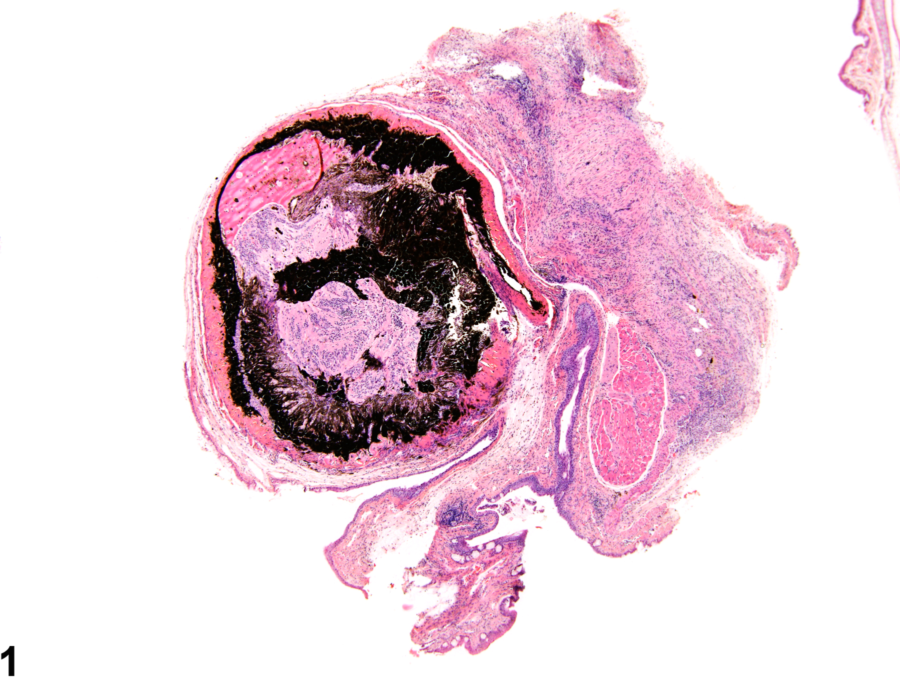
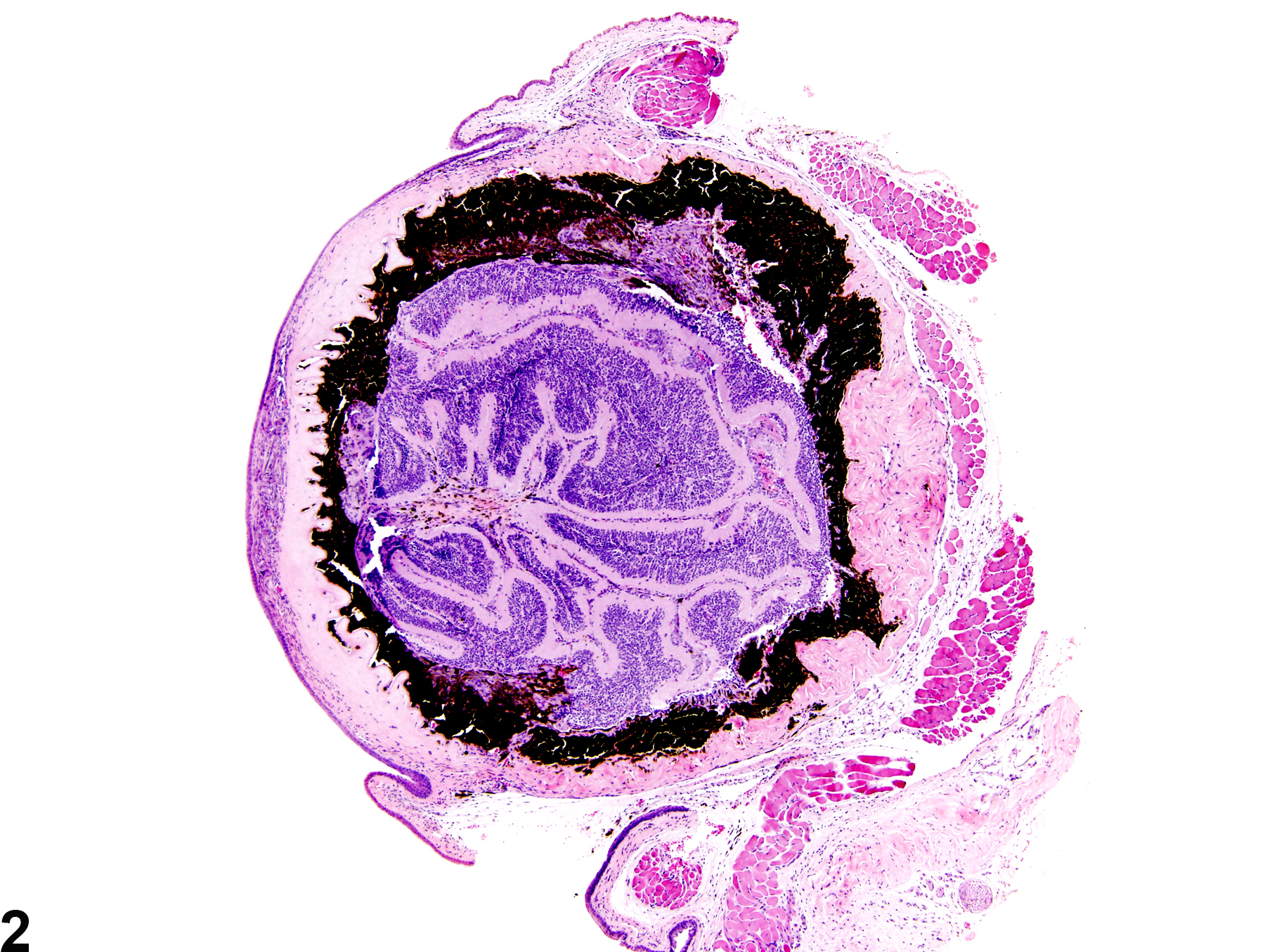
Eye - Phthisis bulbi in a female B6C3F1 mouse from a chronic study. Phthisis bulbi ("end-stage" eye) is characterized by prominent shrinkage of the globe with marked structural displacement and disorganization
All Images

Eye - Phthisis bulbi in a female B6C3F1 mouse from a chronic study. Phthisis bulbi ("end-stage" eye) is characterized by prominent shrinkage of the globe with marked structural displacement and disorganization

Eye - Phthisis bulbi in a female B6C3F1 mouse from a chronic study. The globe is shrunken with marked structural displacement and disorganization.

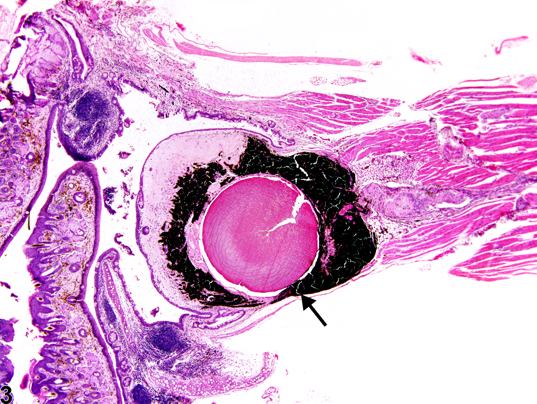
Eye - Phthisis bulbi in a female B6C3F1 mouse from a chronic study. There is marked shrinkage of the globe (arrow) with marked structural displacement and disorganization.

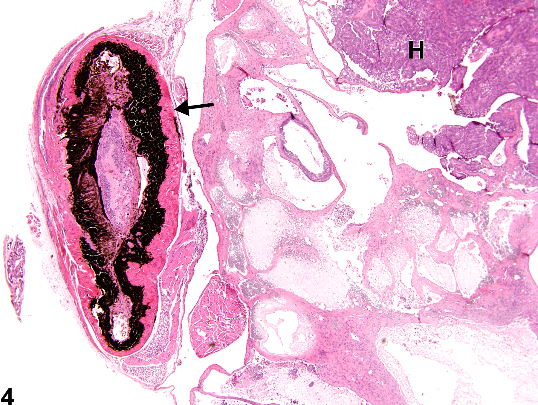
Eye - Phthisis bulbi in a female B6C3F1 mouse from a lifetime study. The globe is shrunken (arrow) with marked structural displacement and disorganization and compressed by a large Harderian gland carcinoma (H).