

Special Senses System
Eye, Vitreous - Proteinaceous Fluid
Narrative
National Toxicology Program. 2012. NTP TR-579. Toxicology and Carcinogenesis Studies of N, N-Dimethyl-p-Toluidine (CAS No. 99-97-8) in F344/N Rats and B6C3F1/N Mice (Gavage Studies). NTP, Research Triangle Park, NC.
Abstract: https://ntp.niehs.nih.gov/go/37162Smith RS. 2002. Choroid, lens, and vitreous. In: Systematic Evaluation of the Mouse Eye: Anatomy, Pathology, and Biomethods (Smith RS, John SWM, Nishina PM, Sundberg JP, eds). CRC Press Boca Raton, FL, 161-193.
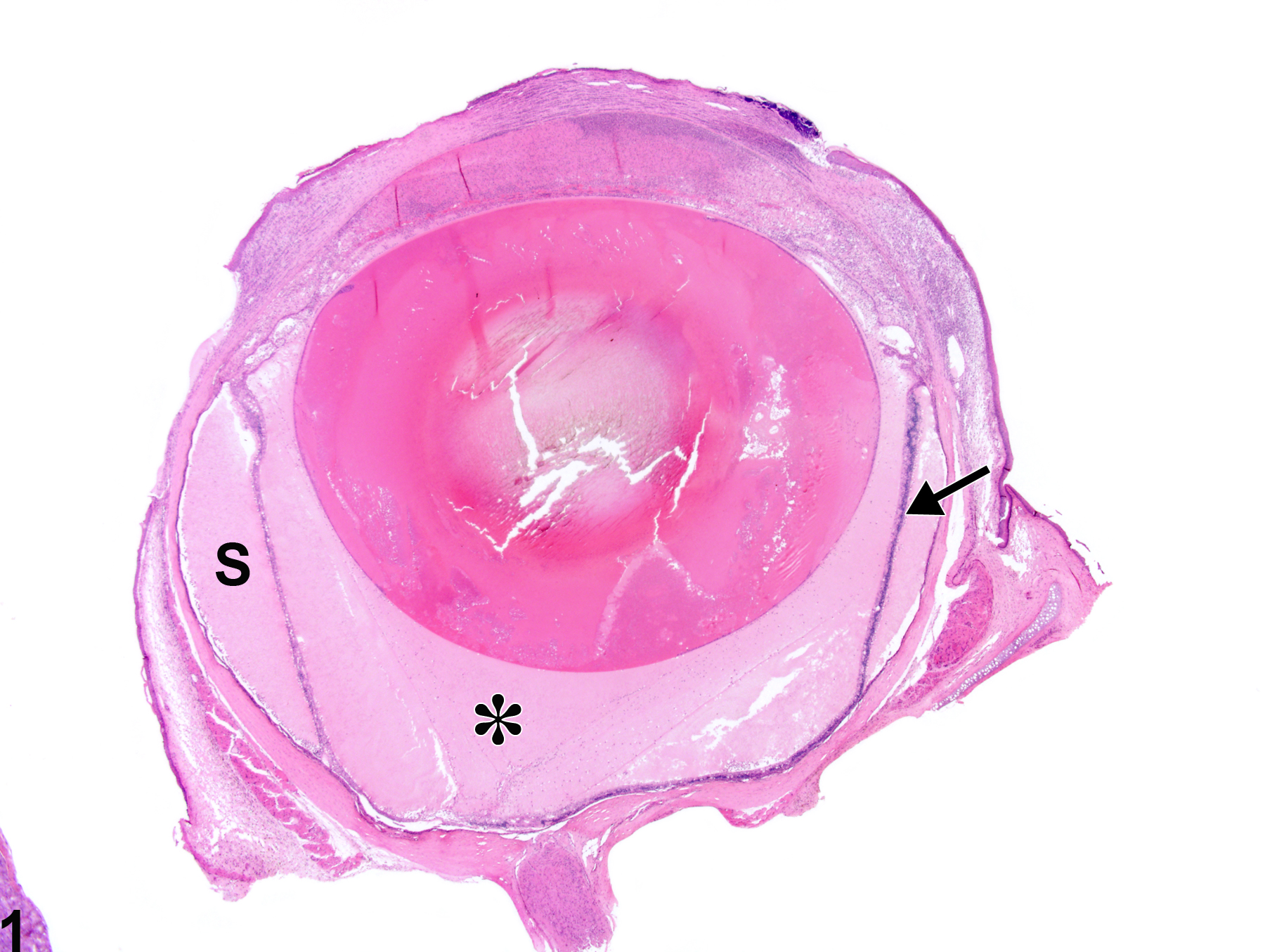
Eye, Vitreous - Proteinaceous fluid in a male F344/N rat from a subchronic study. There is homogeneous pale eosinophilic material (asterisk) with few inflammatory cells posterior to the lens; there is also retinal detachment and degeneration (arrow) and proteinaceous fluid in the subretinal space (S).
All Images

Eye, Vitreous - Proteinaceous fluid in a male F344/N rat from a subchronic study. There is homogeneous pale eosinophilic material (asterisk) with few inflammatory cells posterior to the lens; there is also retinal detachment and degeneration (arrow) and proteinaceous fluid in the subretinal space (S).