

Special Senses System
Harderian Gland - Dilation
Narrative
Botts S, Jokinen M, Gaillard ET, Elwell MR, Mann PC. 1999. Salivary, Harderian, and lacrimal glands. In: Pathology of the Mouse: Reference and Atlas (Maronpot RR, Boorman GA, Gaul BW, eds). Cache River Press, Vienna, IL, 49-79.
Iwai H, Tagawa Y, Hayasaka I, Hayasaka I, Yanai T, Masegi T. 2000. Effects of atropine sulfate on rat Harderian glands: Correlation between morphological changes and porphyrin levels. J Toxicol Sci 25:151-159.
Abstract: http://europepmc.org/abstract/MED/10987121National Toxicology Program. 1993. NTP TR-402. Toxicology and Carcinogenesis Studies of Furan (CAS No. 110-00-9) in F344 Rats and B6C3F1 Mice (Gavage Studies). NTP, Research Triangle Park, NC.
Abstract: https://ntp.niehs.nih.gov/go/12255National Toxicology Program. 1999. NTP TR-469. Toxicology and Carcinogenesis Studies of AZT (CAS No. 30516-87-1) and AZT/α-Interferon A/D in B6C3F1 Mice (Gavage Studies). NTP, Research Triangle Park, NC.
Abstract: https://ntp.niehs.nih.gov/go/6082Satoh Y, Ishikawa K, Oomori Y, Takede S, Ono K. 1992. Secretion mode of the Harderian gland of rats after stimulation by cholinergic secretagogues. Acta Anat 143:7-13.
Abstract: https://www.ncbi.nlm.nih.gov/pubmed/1350161Yoshitomi K, Boorman GA. 1990. Eye and associated glands. In: Pathology of the Fischer Rat: Reference and Atlas (Boorman GA, Eustis SL, Elwell MR, Montgomery CA, MacKenzie WF, eds). Academic Press, San Diego, CA, 239-260.
Abstract: https://www.ncbi.nlm.nih.gov/nlmcatalog/9002563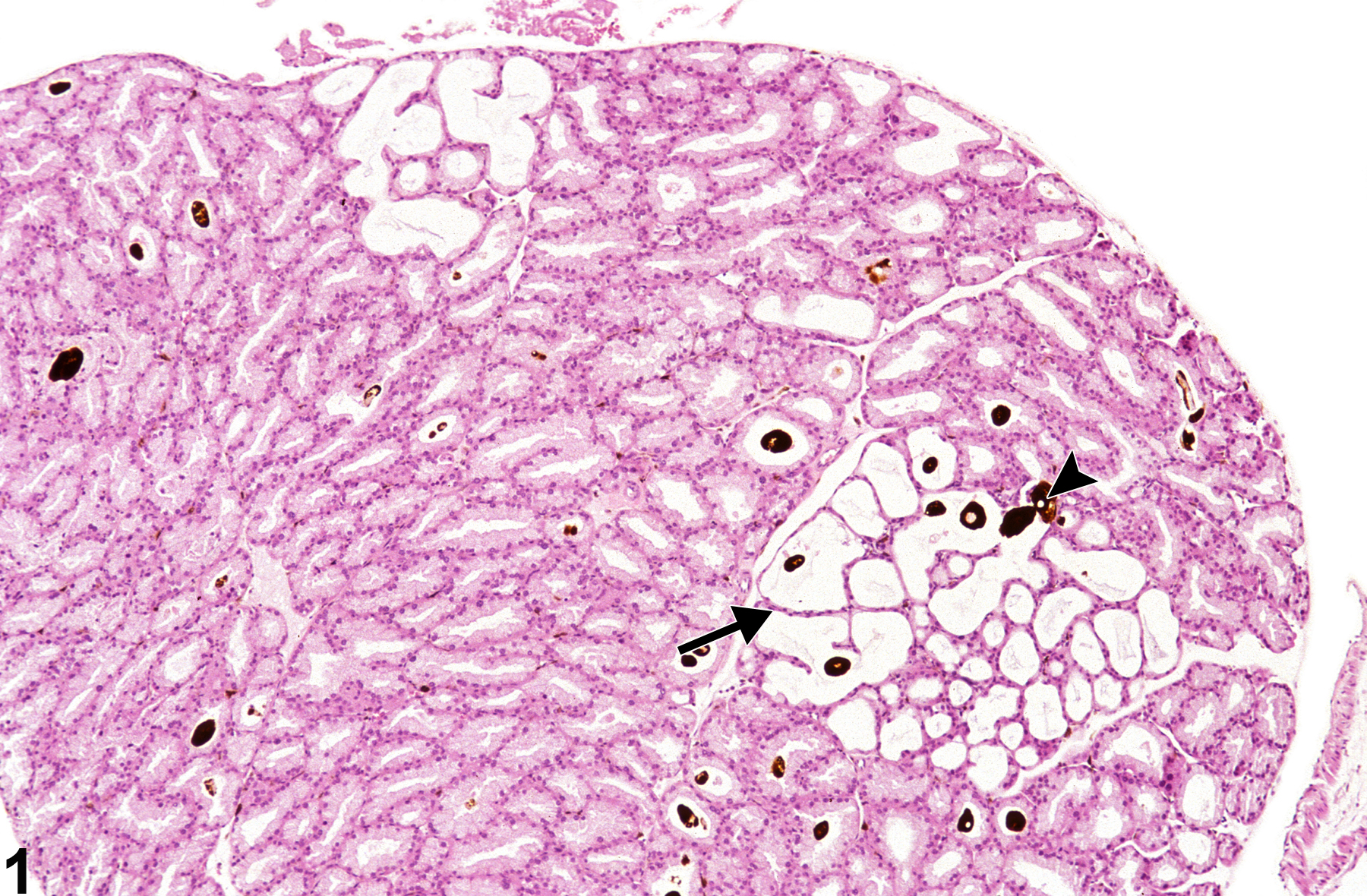
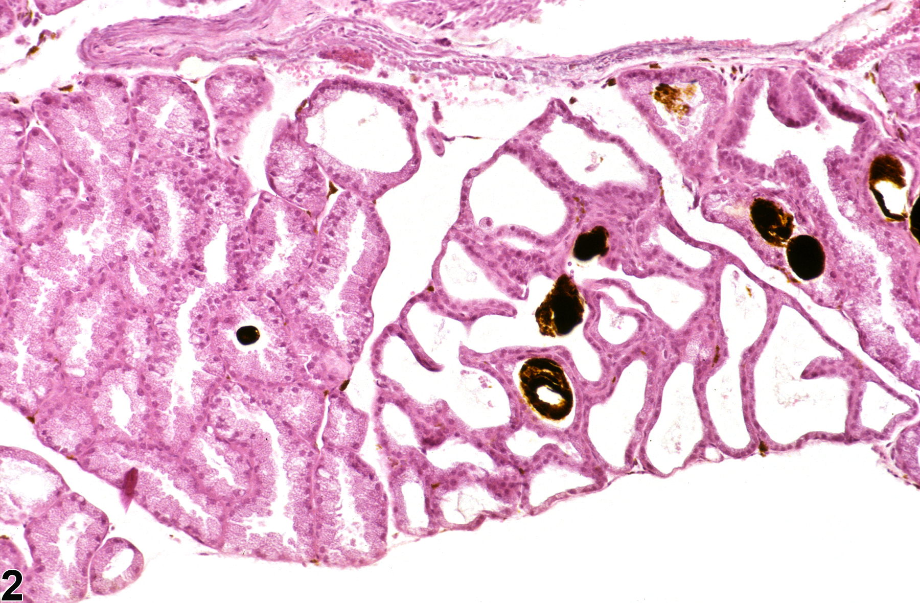
Harderian gland - Dilatation in a male B6C3F1 mouse from a chronic study. There are focal clusters of alveoli with dilated lumens lined by slightly flattened epithelial cells (arrow) some of which contain intraluminal porphyrin-pigment (arrowhead).
All Images

Harderian gland - Dilatation in a male B6C3F1 mouse from a chronic study. There are focal clusters of alveoli with dilated lumens lined by slightly flattened epithelial cells (arrow) some of which contain intraluminal porphyrin-pigment (arrowhead).

Harderian gland - Dilation in a female B6C3F1 mouse from a chronic study. There is little interstitial fibrosis separating the dilated acini.