

Urinary System
Kidney, Papilla, Epithelium - Hyperplasia
Narrative
Frazier KS, Seely JC, Hard GC, Betton G, Burnett R, Nakatsuji S, Nishikawa A, Durchfeld-Meyer B, Bube A. 2012. Proliferative and non-proliferative lesions in the rat and mouse urinary system. Toxicol Pathol 40:14S-86S.
Abstract: http://www.ncbi.nlm.nih.gov/pubmed/22637735McInnes EF. 2012. Wistar and Sprague-Dawley rats. In: Background Lesions in Laboratory Animals: A Color Atlas. Saunders Elsevier, Edinburg, 29.
Abstract: http://www.sciencedirect.com/science/book/9780702035197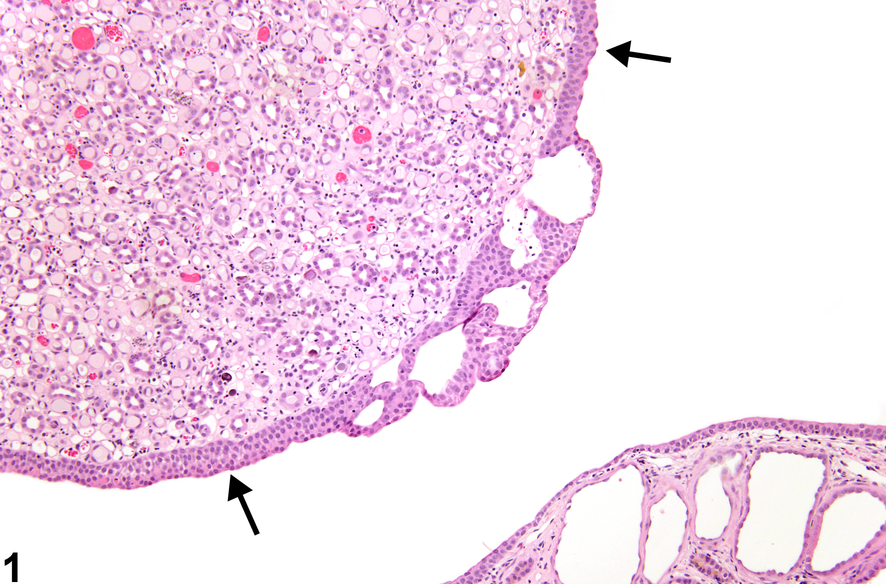
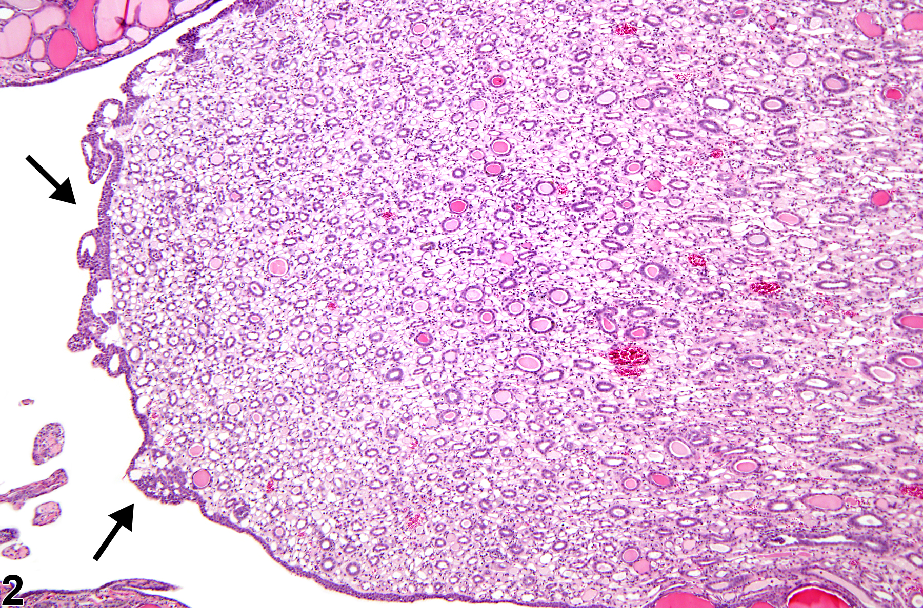
Kidney, Papilla, Epithelium - Hyperplasia in a male F344/N rat from a chronic study. The epithelium is thickened (arrows), and there are large clear spaces within the hyperplastic epithelium.
All Images

Kidney, Papilla, Epithelium - Hyperplasia in a male F344/N rat from a chronic study. The epithelium is thickened (arrows), and there are large clear spaces within the hyperplastic epithelium.

Kidney, Papilla, Epithelium - Hyperplasia in a male F344/N rat from a chronic study. A small focus of hyperplastic surface epithelial cells lines the renal papilla (arrows).