

Integumentary System
Mammary Gland - Acinar Atrophy
Narrative
Comment:
Diffuse atrophy of the normal alveolar (acinar) (Figure 1, Figure 2, Figure 3, and Figure 4) and ductular (Figure 3 and Figure 4) tissue of the mammary gland of rats and mice is an uncommon finding. It is most often associated with compound administration. Involution of the mammary gland begins in the second year of life in rats and mice and is characterized by loss of distal ducts in mice, and regression of terminal end buds and decrease in size of lobules in rats. This change, of course, would be present in both treated and control animals. Treatment with certain xenobiotics for long periods can induce hormonal perturbations, such as hypoestrogenism or estrogen receptor (ER) antagonism in rats that causes atrophy of the female mammary gland. With atrophy of the female mammary glands, the ductular and acinar components are both affected, resulting in a prominent appearance of the mammary gland fat pad. The epithelial cells lining the affected ducts and acini are low cuboidal and have a high nuclear cytoplasmic ratio. The term feminization refers to the conversion of the normally lobuloalveolar pattern of male rat mammary glands to the tubuloalveolar pattern seen in female rats and thus differs slightly from atrophy. Tamoxifen and toremifene are selective estrogen receptor modulators (SERM) and are examples of potent estrogen receptor antagonists in the rat mammary gland that cause ductal atrophy (and ectasia) in females and acinar atrophy in males. Care must be taken during tissue sectioning to prevent the erroneous appearance of atrophy during histopathologic evaluation.
Recommendations:
Atrophy of the mammary gland should be diagnosed and graded in males and females whenever present. The appropriate topographic modifier (“duct” or “acinus”) should be included in the diagnosis. If atrophy of both the ducts and acini are present, the topographic modifier may be omitted and the affected structures described in the pathology narrative. The term “feminization” of the mammary gland is not recommended because it is indicative of a syndrome that only applies to male rats. Involution of the mammary glands in older animals should not be diagnosed as atrophy.
References:
| Boorman GA, Wilson JT, Van Zwieten M, Eustis SL. 1990. Mammary gland. In: Boorman GA, Eustis SL, Elwell MR, Montgomery CA, Mackenzie WF (eds.). 2016. Pathology of the Fischer rat - reference and atlas. Academic Press pp. 295-313. |
| Greaves P. 2007. Mammary gland. Histopathology of preclinical toxicity studies. Interpretation and relevance in drug safety evaluation, 3rd ed. Academic Press pp. 68-98. |
| Greaves P, Goonetilleke R, Nunn G, Topham J, Orton T. 1993. Two-year carcinogenicity study of tamoxifen in Alderley Park Wistar-derived rats. Cancer Res 53(17):3919-24. |
| Lucas JN, Rudmann DG, Credille KM, Irizarry AR, Peter A, Snyder PW. 2007. The rat mammary gland: morphologic changes as an indicator of systemic hormonal perturbations induced by xenobiotics. Toxicol Pathol 35:199-207. |
| Kennel P, Pallen C, Barale-Thomas E, Espuna G, Bars R. 2003. Tamoxifen: 28-day oral toxicity study in the rat based on the enhanced OECD test guideline 407 to detect endocrine effects. Arch Toxicol 77(9):487-99. |
| Richert MM, Schwertfeger KL, Ryder JW, Anderson SM. 2000. An atlas of mouse mammary gland development. J Mammary Gland Biol Neoplasia 5(2):227-41. |
Seely JC, Boorman GA. 1999. Mammary gland and specialized sebaceous glands. In: Pathology of the Mouse: Reference and Atlas (Maronpot RR, Boorman GA, Gaul BW, eds). Cache River Press, Vienna, IL, 613-635.
| Sourla A, Martel C, Labrie C, Labrie F. 1998. Almost exclusive androgenic action of dehydroepiandrosterone in the rat mammary gland. Endocrinology 139(2):753-64. Abstract: https://pubmed.ncbi.nlm.nih.gov/9449650 |
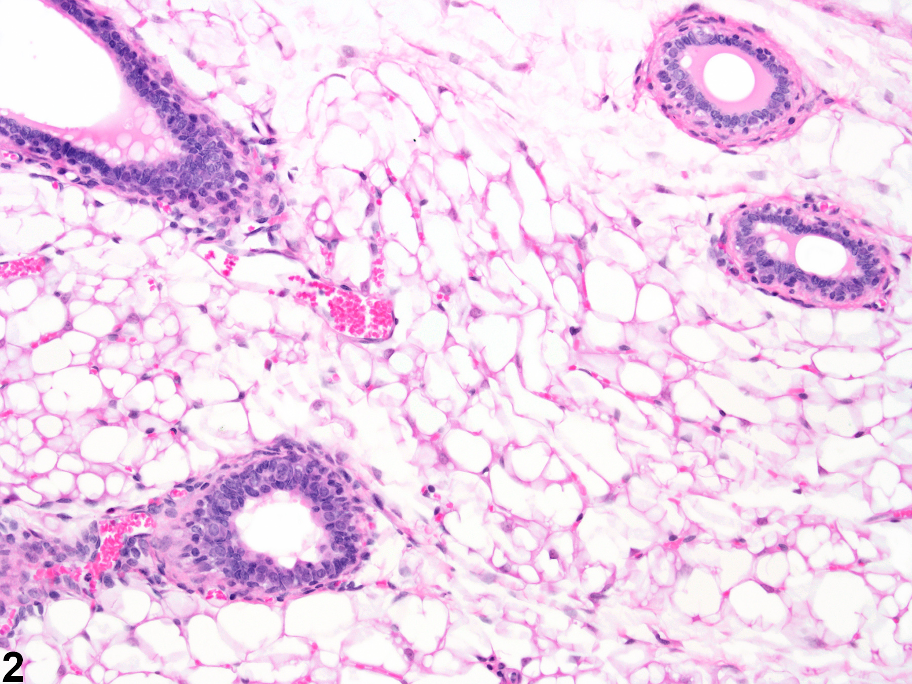 Mammary gland - Normal in a female B6C3F1/N mouse from an acute study (higher magnification of Figure 1). Normal mammary gland with well-developed mammary fat (adipose) pad, ductules, and acini.
All Images
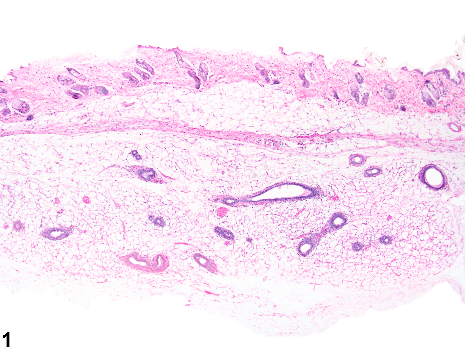
Mammary gland - Normal in a female B6C3F1/N mouse from an acute study. Normal mammary gland with well-developed mammary fat (adipose) pad and age appropriate ductular and acinar tissue.

Mammary gland - Normal in a female B6C3F1/N mouse from an acute study (higher magnification of Figure 1). Normal mammary gland with well-developed mammary fat (adipose) pad, ductules, and acini.
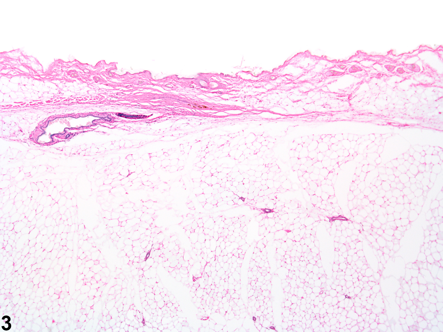
Mammary gland, Acinus - Atrophy in a female B6C3F1/N mouse from an acute study. There is diffuse depletion of the ductules and acinar tissue with a normal well-developed fat pad (compare to Figure 1).
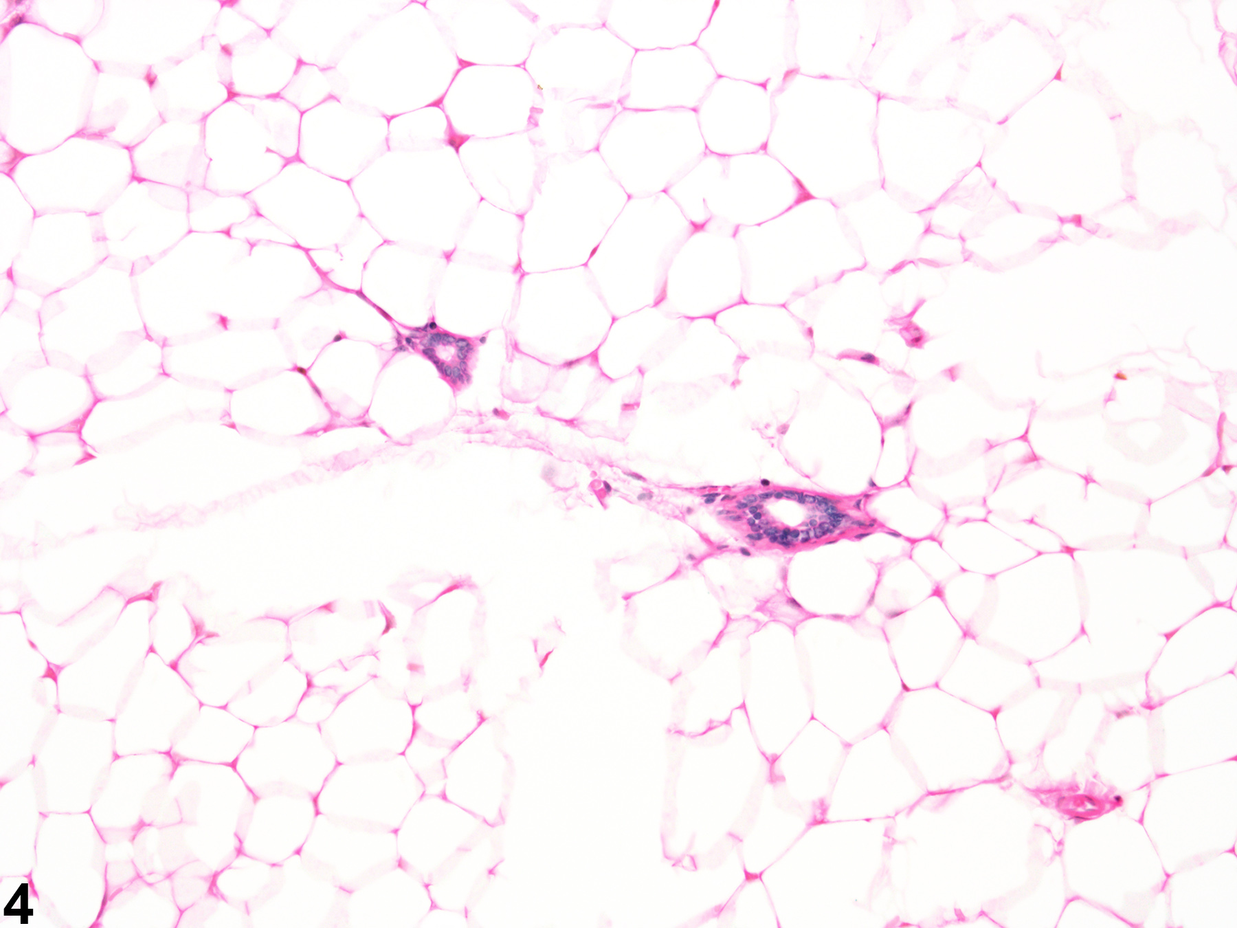
Mammary gland, Acinus - Atrophy in a female B6C3F1/N mouse from an acute study (higher magnification of Figure 3). The acini are decreased in size (compare to Figure 2).