Integumentary System
Mammary Gland - Galactocele
Narrative
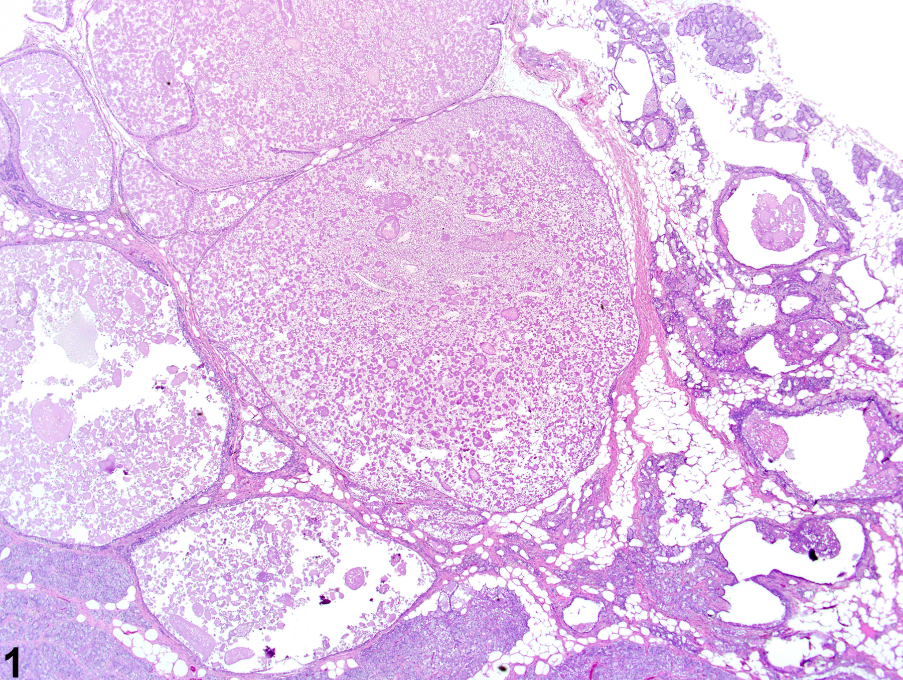
Mammary gland galactoceles are markedly dilated/cystic ducts and alveoli filled with proteinaceous secretory material, which are generally detectable grossly at necropsy. Galactoceles are considered a form of ductular or acinar dilation, which is often diffuse and characterized by distention of collecting (lactiferous) ducts and alveoli, often beneath a nipple, and accompany additional histological changes other than just distension. Galactoceles are typically lined by flattened to cuboidal epithelium, which may be vacuolated, with a thin connective tissue wall and filled with proteinaceous secretory fluid and cell debris. Galactoceles may be accompanied by necrotic debris, inflammation, and thickened fibrotic walls, which are thought to be secondary to leakage or rupture.
Galactoceles may occur as a spontaneous age-related change or can be caused by anything that blocks an outlet (e.g., lactiferous duct occlusion during lactation or in response to hyperprolactinemia). In rodents, galactoceles are commonly associated with mammary gland neoplasms that cause blockage of a secretory duct.
| Boorman GA, Wilson JT, Van Zwieten M, Eustis SL. 1990. Mammary gland. In: Boorman GA, Eustis SL, Elwell MR, Montgomery CA, Mackenzie WF (eds.). 2016. Pathology of the Fischer rat - reference and atlas. Academic Press pp. 295-313. |
| Burek JD. 1978. Pathology of aging rats. CRC Press pp. 163-167. |
| Yi ES, Bedoya AA, Lee H, Kim S, Housley RM, Aukerman SL, Tarpley JE, Starnes C, Yin S, Pigrce GF, Ulich TR. 1994. Keratinocyte growth factor causes cystic dilatation of the mammary Glands of mice. Am J Pathol 145(5):1015-1022. |
| Goodman DG, Ward JM, Squire RA, Chu KC, Linhart MS. 1979. Neoplastic and nonneoplastic lesions in aging F344 rats. Toxicol Appl Pharmacol 48(2):237-248. |
| Greaves P. 2007. Mammary gland. Histopathology of preclinical toxicity studies. Interpretation and relevance in drug safety evaluation, 3rd ed. Academic Press pp. 68-98. |
| Lee K-Y, Shibutani M, Takagi H, Kato N, Takigami S, Uneyama C, Hirose M. 2004. Diverse developmental toxicity of di-n-butyl phthalate in both sexes of rat offspring after maternal exposure during the period from late gestation through lactation. Toxicology 203(1-3):221-38. |
| Rehm S, Liebalt AG. Nonneoplastic and neoplastic lesions of the mammary gland. In: Mohr U, Dungworth DL, Ward J, Capen CC, Carlton WW, Sundberg JP (eds.). 1996. Pathobiology of the aging mouse, Vol. 2. International Life Sciences Institute Press pp. 381-398. |
| Russo J, Russo IH, Van Zwieten MJ, Rogers AE, Gusterson BA. Classification of neoplastic and nonneoplastic lesions of the rat mammary gland. In: Jones TC, Mohr U, Hunt RD (eds.). 1989. Integument and mammary glands (monographs on pathology of laboratory animals). International Life Sciences Institute pp. 275-304. |
| Seely JC, Boorman GA. Mammary gland and specialized sebaceous glands. In: Maronpot RR, Boorman GA, Gaul BW (eds.). 1999. Pathology of the mouse: reference and atlas. Cache River Press pp. 613-635. |
| Tucker MJ. 1997. The integumentary system and mammary glands. Diseases of the Wistar rat. CRC Press pp. 23-36. |
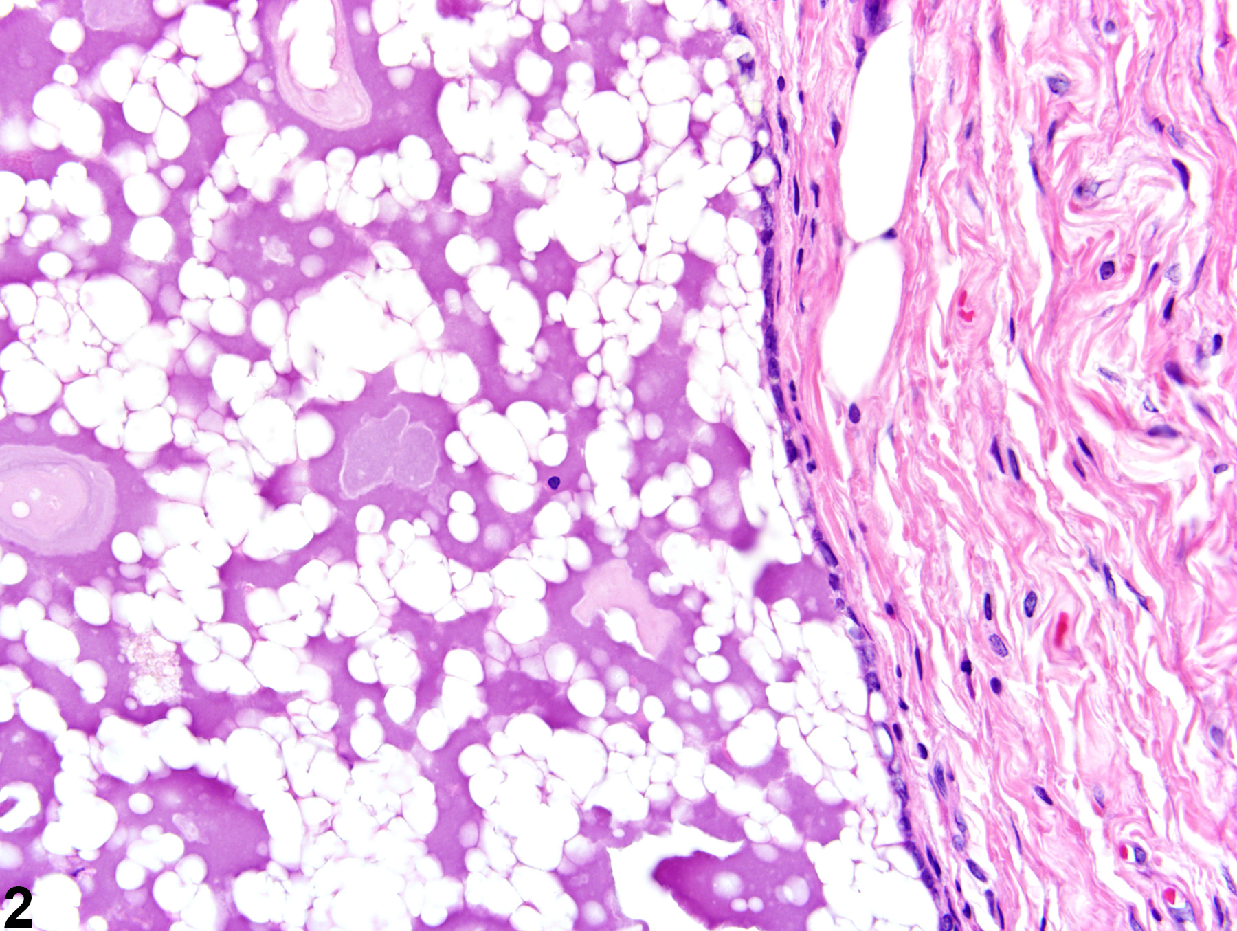 Mammary gland - Galactocele in a female F344/N rat from a chronic study (higher magnification of Figure 1). A distended alveolus contains eosinophilic secretory material, and the epithelial lining is flattened and vacuolated.
All Images